Opening Hours : Mon to Sat - 9am to 9pm / Sun - Closed

Diphtheria

Definition of Diphtheria:

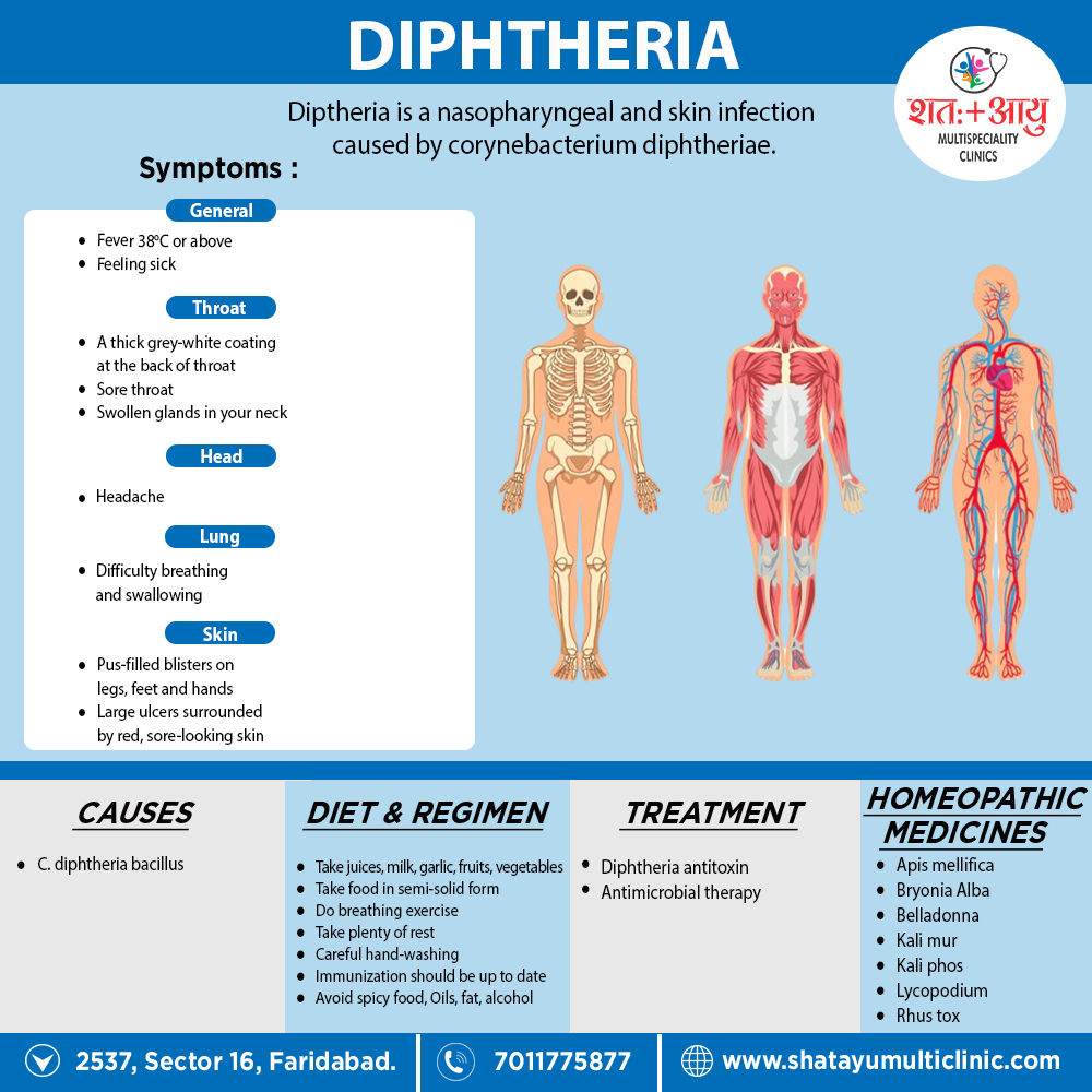
Diphtheria is a nasopharyngeal and skin infection caused by Corynebacterium diphtheriae. Toxigenic strains of C. diphtheriae produce a protein toxin that causes systemic toxicity, myocarditis, and polyneuropathy. The toxin is associated with the formation of pseudomembranes in the pharynx during respiratory diphtheria. While toxigenic strains most frequently cause pharyngeal diphtheria, nontoxigenic strains commonly cause cutaneous disease. [1]

Overview

Overview of diphtheria

While in many regions diphtheria has been controlled in recent years with effective vaccination, there have been sporadic outbreaks in the United States and Europe. Diphtheria is still common in the Caribbean, Latin America, and the Indian subcontinent, where mass immunization programs are not enforced. Largescale epidemics of diphtheria have occurred in the post-Soviet independent states. Additional outbreaks have been reported in Algeria, China, and Ecuador.

History of diphtheria

The development of diphtheria antitoxin in 1898 by von Behring also of the diphtheria toxoid vaccine in 1924 by Ramon led to the near elimination of diphtheria in Western countries. The annual incidence rate in the United States peaked in 1921 at 191 cases per 100,000 populations. In contrast, since 1980, the annual figure in the United States has been. Nevertheless, pockets of colonization persist in North America, particularly in South Dakota, Ontario, and recently the state of Washington. Immunity to diphtheria induced by childhood vaccination gradually decreases in adulthood. In detail; An estimated 30% of men 60–69 years old have antitoxin titers below the protective level. In addition to older age and lack of vaccination, risk factors for diphtheria outbreaks include alcoholism, low socioeconomic status, crowded living conditions, and Native American ethnic background.

Clonally related toxigenic C. diphtheriae strains of the ET8 complex associate with this outbreak. Given that the ET8 complex expressed a toxin against which the prevalent diphtheria toxoid vaccine was effective, the epidemic attribute to failure of the public health infrastructure to effectively vaccinate the population. Socioeconomic instability, migration, deteriorating public health programs, frequent vaccine shortages, delayed implementation of vaccination and treatment in response to cases, also lack of public education and awareness were contributing factors. [1]

Causes

Diphtheriae is a gram-positive bacillus that unencapsulated, nonmotile, also nonsporulating. The organism first identified microscopically in 1883 by Klebs and a year later isolated in pure culture by Löffler in Robert Koch’s laboratory. The bacteria have a characteristic club-shaped bacillary appearance and typically form clusters of parallel rays, or palisades, that referred to as “Chinese characters.”

Different Laboratory media

The specific laboratory media recommended for the cultivation of C. diphtheriae rely upon tellurite, colistin, or nalidixic acid for the organism’s selective isolation from other autochthonous pharyngeal microbes. C. diphtheriae may isolated from individuals with both nontoxigenic (tox–) and toxigenic (tox+) phenotypes.

Uchida and Pappenheimer demonstrated that coryne-bacteriophage beta carries the structural gene tox, which encodes diphtheria toxin, also that a family of closely related corynebacteriophages are responsible for toxigenic conversion of tox– C. diphtheriae to the tox+ phenotype. Moreover, lysogenic conversion from a nontoxigenic to a toxigenic phenotype has shown to occur in situ. Growth of toxigenic strains of C. diphtheriae under iron-limiting conditions leads to the optimal expression of diphtheria toxin and believed to a pathogenic mechanism during human infection [1]

Pathophysiology

Diphtheria toxin produced by tox+ strains of C. diphtheriae is the primary virulence factor in clinical disease. The toxin synthesize in precursor form; released as a 535-amino-acid, single-chain protein; also , in sensitive species (e.g., guinea pigs and humans, but not mice or rats), has a 50% lethal dose of ~100 ng/kg of body weight. The toxin produce in the pseudomembranous lesion and taken up in the bloodstream, from which it distribute to all organ systems in the body. Once bound to its cell surface receptor (e.g. a heparin-binding epidermal growth factor–like precursor), the toxin internalized by receptor-mediated endocytosis and enters the cytosol from an acidified early endosomal compartment.

In vitro, the toxin may separated into two chains by digestion with serine proteases: the N-terminal A fragment and the C-terminal B fragment. Delivery of the A fragment into the eukaryotic cell cytosol results in irreversible inhibition of protein synthesis by NAD+-dependent ADP-ribosylation of elongation factor 2. The eventual result is the death of the cell. [1]

Sign & Symptoms

Symptoms of Diphtheria:

Symptoms usually start 2 to 5 days after becoming infected.

The main symptoms of diphtheria are:

  • A thick grey-white coating at the back of your throat
  • A high temperature (in other words; fever) of 38C or above
  • Feeling sick
  • Sore throat
  • Headache
  • Swollen glands in your neck
  • Difficulty breathing and swallowing

If it affects your skin (in other words; cutaneous diphtheria), it can cause:

  • pus-filled blisters on your legs, feet and hands
  • large ulcers surrounded by red, sore-looking skin [1]

Diagnosis

The diagnosis of diphtheria is based on clinical signs and symptoms plus laboratory confirmation. Respiratory diphtheria should considered in patients with sore throat, pharyngeal exudates, and fever. Other symptoms may include hoarseness, stridor, or palatal paralysis. The presence of a pseudomembrane should prompt strong consideration of diphtheria. Once a clinical diagnosis of diphtheria made, diphtheria antitoxin should obtained and administered as rapidly as possible.

Laboratory diagnosis

Laboratory diagnosis of diphtheria based either on cultivation of C. diphtheriae or toxigenic Corynebacterium ulcerans from the site of infection or on the demonstration of local lesions with characteristic histopathology. Corynebacterium pseudodiphtheriticum, a nontoxigenic organism, is a common component of the normal throat flora and does not pose a significant risk. Throat samples should submit to the laboratory for culture with the notation that diphtheria is being considered. This information should prompt cultivation on special selective medium and subsequent biochemical testing to differentiate C. diphtheriae from other nasopharyngeal commensal corynebacteria. All laboratory isolates of C. diphtheriae, including nontoxigenic strains, should submit to the CDC.

Cutaneous diphtheria

A diagnosis of cutaneous diphtheria requires laboratory confirmation since the lesions are not characteristic and are indistinguishable from other dermatoses. Diphtheritic ulcers occasionally—but not consistently—have a punched-out appearance (Fig. 175-2). Patients in whom cutaneous diphtheria identified should have the nasopharynx cultured for C. diphtheriae. The laboratory medium for cutaneous diphtheria specimens the same as that used for respiratory diphtheria: Löffler’s or Tinsdale’s selective medium in addition to nonselective medium such as blood agar. As has mentioned, respiratory diphtheria remains a notifiable disease in the United States, whereas cutaneous diphtheria is not. [3]

Investigation of Diphtheria:

  1. diphtheriae transmitted via the aerosol route, usually during close contact with an infected person. There are no significant reservoirs other than humans. The incubation period for respiratory diphtheria is 2–5 days, but disease onset has occurred as late as 10 days after exposure. Prior to the vaccination era, most individuals over the age of 10 were immune to C. diphtheriae; infants protected by maternal IgG antibodies but became susceptible after ~6 months of age. Thus, the disease primarily affected children and nonimmune young adults. In temperate regions, respiratory diphtheria occurs year-round but is most common during winter months [1]

Treatment

Treatment

DIPHTHERIA  ANTITOXIN

Prompt administration of diphtheria antitoxin is critical in the management of respiratory diphtheria. Diphtheria antitoxin, a horse antiserum, is effective in reducing the extent of local disease as well as the risk of complications of myocarditis and neuropathy. Rapid institution of antitoxin therapy also associated with a significant reduction in mortality risk. Because diphtheria antitoxin cannot neutralize cell-bound toxin, prompt initiation is important. This product, which is no longer commercially available in the United States, can obtain from the CDC by calling the Bacterial Vaccine Preventable Disease Branch of the National Immunization Program at 404-639-8257 (8:00 a.m. to 4:30 p.m., U.S. Eastern time) or, at other hours, the Emergency Operations Center at 770-488-7100; the relevant website is www.cdc.gov/diphtheria/dat.html. The current protocol for the use of diphtheria antitoxin involves a test dose to rule out immediate hypersensitivity. Patients who demonstrate hypersensitivity require desensitization before a full therapeutic dose of antitoxin administered

ANTIMICROBIAL THERAPY

Antibiotics used in the management of diphtheria primarily to prevent transmission to susceptible contacts. Antibiotics also prevent further toxin production and reduce the severity of local infection. Recommended treatment options for patients with respiratory diphtheria are as follows:

  • Procaine penicillin G, 600,000 U IM q12h (for children: 12,500–25,000 U/kg IM q12h) until the patient can swallow comfortably; then oral penicillin V, 125–250 mg qid to complete a 14-day course
  • Erythromycin, 500 mg IV q6h (for children: 40–50 mg/kg per day IV in two or four divided doses) until the patient can swallow comfortably; then 500 mg PO qid to complete a 14-day course A clinical study in Vietnam found that penicillin associate with a more rapid resolution of fever and a lower rate of bacterial resistance than erythromycin; however, relapses were more common in the penicillin group. Erythromycin therapy targets protein synthesis and thus offers the presumed benefit of stopping toxin synthesis more quickly than a cell wall– active β-lactam agent. Alternative therapeutic agents for patients who are allergic to penicillin or cannot take erythromycin include rifampin and clindamycin. Eradication of C. diphtheriae should document after antimicrobial therapy is complete.
Throat culture

A repeat throat culture 2 weeks later is recommended. For patients in whom the organism is not eradicating after a 14-day course of erythromycin or penicillin, an additional 10-day course followed by repeat culture is recommended. Drug-resistant strains of C. diphtheriae exist, and several reports have described multidrug resistant strains, predominantly in Southeast Asia. Drug resistance should consider when efforts at pathogen eradication fail. Cutaneous diphtheria should treated as described above for respiratory disease. Individuals infected with toxigenic strains should receive antitoxin. It is important to treat the underlying cause of the dermatoses in addition to the superinfection with C. diphtheriae. Patients who recover from respiratory or cutaneous diphtheria should have antitoxin levels measured. If diphtheria antitoxin has administered, this test should performed 6 months later. Patients who recover from respiratory or cutaneous diphtheria should receive the appropriate vaccine to ensure the development of protective antibody titers. [1]

MANAGEMENT STRATEGIES:

Patients in whom diphtheria is suspected should hospitalized in respiratory isolation rooms, with close monitoring of cardiac and respiratory function. A cardiac workup recommended to assess the possibility of myocarditis. In patients with extensive pseudomembranous, an anesthesiology or an ear, nose, and throat consultation recommended because of the possible need for tracheotomy or intubation. In some settings, pseudomembranous can remove surgically. Treatment with glucocorticoids has not been shown to reduce the risk of myocarditis or polyneuropathy. [1]

Homeopathic Treatment

Homeopathic medicines are individualized, selected based on the individual case history of the patient, by taking into consideration the cause like hormonal imbalance, nutritional factor, emotional stress, the nature of the patient, and other factors which may be acting as a maintaining cause in the diphtheria.

Diphtheria, as it is an acute problem, requires a constitutional treatment. Homeopathy focuses on the root cause of the problem and helps in the treatment of diphtheria.

Medicines:

  1. Bryonia Alba:

Great prostration; patient cannot bear to be moved; complains of pains everywhere when moved; white tongue; dry mouth; thirst for large quantities of water.

  1. Belladona:

Restless; complains of sore throat, which looks highly inflamed; pupils large; drowsy, but unable to sleep; starts suddenly out of sleep

  1. Lycopodium:

Throat brownish red; worse right side; worse from swallowing warm drinks; nose stopped up. Furthermore; patient cannot breathe with mouth shut; keeps mouth constantly open, slightly protruding the tongue, which gives a silly expression; unsteady step; on awaking from short nap he is cross; kicks; is naughty (if a child); or jumps up in bed, stares about, also knows nobody seemingly dreaming with open eyes; frequent jerkings of lower limbs, mostly with a groan, awake or slumbering; great fear of being left alone.

  1. Rhus
    tox:

Restless; wants to be carried about; wakes complaining of pain in throat; bloody saliva runs out of mouth during sleep; parotid glands good deal swollen; transparent jelly-like discharges form bowels at stool or afterwards

  1. Apis mellifica:

Great debility from the beginning; membranes at once assume dirty greyish colour, or there is great oedema of soft palate and much puffiness round the eyes; pain in ears on swallowing; an itching, stinging eruption on skin; sensation of weakness in larynx; numbness of feet and hands and even paralysis. (Apis is incompatible with Rhus, and should not be given immediately after it or Rhus after Apis.)

  1. Kali Mur: For typical diphtheria.
  2. Kali phos:

When the breath becomes putrid and the throat gangrenous or the case paralytic.

  1. Ammonium carb:

Diphtheria of scrofulous persons with swelling of the glands of the neck; putrid sore throat, burning pain with roughness also scraping in throat; tendency to gangrene in throat. Additionally, nose obstructed, child aroused from sleep by want of breath; great prostration; excessive sensitiveness to cold air and cold drinks; Besides this; cerebral symptoms

  1. Arsenicum iod:

Diphtheritic croup, deposit covering mouth from fauces to outer edge of lips and external auditory canal; foul breath; short, difficult respiration and symptoms of adynamia marked enlargement of lymphatic glands, WEAK HEART.

10. Bromium:

Malignant forms of diphtheria, invading LARYNX DOWN RESPIRATORY ORGANS, leaving great weakness and lassitude; no fever, cool skin, sweating and spasm; husky tone of voice, rattling of mucus in larynx when coughing , cough has a croupy sound, with strangling of mucus in larynx when coughing rattling mucus in breathing; face ashy-gray, cheeks sunken, stiffness of neck, prostration Suits children with fair hair and skin, blue eyes, etc., also Teste affirms that milk neutralizes the action of Brom. And Iod. (Kali bi. no spasm). [4]

Diet & Regimen

Foods to be taken

  • Fluids (e.g. juices)
  • Milk
  • Garlic
  • Food in semi-solid form (e.g. ice creams)
  • Fruits and vegetables

Foods to be avoided

  • Spicy food
  • Oils and fats
  • Alcohol

Yoga and Exercise

  • Breathing exercise (e.g. pranayama)
  • Uttanasana
  • Sarvangasana

Home Remedies

  • Drink plenty of water
  • Take plenty of rest
  • Careful hand-washing
  • Take diet through liquids and soft foods
  • Immunization should be up to date
  • Patient should avoid sleeping on their back to steer clear of swallowing the secretion formed in the mouth [2]

FAQs

Frequently Asked Questions

What is Diphtheria?

Diphtheria is a nasopharyngeal and skin infection caused by Corynebacterium diphtheriae. Toxigenic strains of C. diphtheriae produce a protein toxin that causes systemic toxicity, myocarditis, and polyneuropathy

Homeopathic Medicines used by Homeopathic Doctors in treatment of Diphtheria?

  • Bryonia Alba
  • Belladona
  • Lycopodium
  • Rhus tox
  • Apis mellifica
  • Kali Mur
  • Arsenicum iod
  • Bromium

What causes Diphtheria?

Corynebacterium diphtheriae gram-positive bacillus

What are the symptoms of Diphtheria?

  • A thick grey-white coating at the back of your throat
  • A high temperature (fever) of 38C or above
  • Feeling sick
  • Sore throat
  • Headache
  • Swollen glands in your neck
  • Difficulty breathing and swallowing

References:

  1. Harrison-s_Principles_of_Internal_Medicine-_19th_Edition-_2_Volume_Set
  2. https://www.mtatva.com/en/disease/diphtheria-treatment-diet-and-home-remedies/
  3. https://www.nhs.uk/conditions/diphtheria/
  4. A Dictionary Of Domestic Medicine By John H.Clarke.