Opening Hours : Mon to Sat - 9am to 9pm / Sun - Closed

Chronic Otitis Media

Definition:

Chronic suppurative otitis media (CSOM) is a chronic inflammation of the middle ear and mastoid cavity that is characterize by discharge from the middle ear through a perforated tympanic membrane for at least 6 weeks. [1]

Overview

Incidence of CSOM is higher in developing countries because of poor socioeconomic standards, poor nutrition and lack of health education. It affects both sexes and all age groups. In India, the overall prevalence rate is 46 and 16 persons per thousand in rural and urban population, respectively. It is also the single most important cause of hearing impairment in rural population.

CSOM occurs following an upper respiratory tract infection that has led to acute otitis media. This progresses to a prolonged inflammatory response causing mucosal (middle ear) oedema, ulceration and perforation. The middle ear attempts to resolve this ulceration by production of granulation tissue and polyp formation. This lead to increase discharge and failure to arrest the inflammation, and to development of CSOM, which is also often associated with cholesteatoma. There may be enough pus that it drains to the outside of the ear (in other words, otorrhea), or the pus may be minimal enough to be seen only on examination with an otoscope or binocular microscope. Hearing impairment often accompanies this disease.[1][2]

Types

Clinically, it is divided into two types:

1.Tubotympanic.

Also called the safe or benign type; it involves anteroinferior part of middle ear cleft, i.e., eustachian tube and mesotympanum and is associated with a central perforation. There is no risk of serious complications.

2.Atticoantral.

It involves posterosuperior part of middle ear cleft (attic, antrum, posterior tympanum and mastoid) and is associated with cholesteatoma, which, because of its bone eroding properties, causes risk of serious complications. For this reason, the disease is also called unsafe or dangerous type.[2]

Causes

[1] Tubotympanic

The disease starts in childhood and is therefore common in that age group.

1.  It is the sequela of acute otitis media usually following exanthemata’s fever and leaving behind a large central perforation. The perforation becomes permanent and permits repeated infection from the external ear. Also, the middle ear mucosa is exposed to the environment and gets sensitized to dust, pollen and other aeroallergens causing persistent otorrhea.

2. Ascending infections via the eustachian tube. Infection From tonsils, adenoids and infected sinuses may be responsible for persistent or recurring otorrhoea. Ascending infection to middle ear occurs more easily in the presence of infection.

3. Persistent mucoid otorrhoea is sometimes the result of allergy to milk, eggs, fish, etc.

[2] Atticoantral.

Aetiology of atticoantral disease is same as of cholesteatoma and has been discussed earlier. It is seen in sclerotic mastoid, and whether the latter is the cause or effect of disease is not yet clear.[2]

Pathophysiology

A. Tubotympanic.

The tubotympanic disease remains localized to the mucosa and, that too, mostly to anteroinferior part of the middle ear cleft. Like any other chronic infection, the processes of healing and destruction go hand in hand and either of them may take advantage over the other, depending on the virulence of organism and resistance of the patient. Thus, acute exacerbations are not uncommon. The pathological changes seen in this type of CSOM are:

1.Perforation of pars tensa. It is a central perforation and its size and position vary.

2.Middle ear mucosa. It may be normal when disease is quiescent or inactive. It is oedematous and velvety when disease is active.

3.Polyp. A polyp is a smooth mass of oedematous and inflamed mucosa which has protruded through a perforation and presents in the external canal. It Is usually pale in contrast to pink, fleshy polyp seen in attic-antral disease.[2]

4.Ossicular chain. It Is usually intact and mobile but may show some degree of necrosis, particularly of the long process of incus.

5. Tympanosclerosis which shown by hyalinization and subsequent calcification of subepithelial connective tissue. It is seen in remnants of tympanic membrane or under the mucosa of middle ear. It is seen as white chalky deposit on the promontory, ossicles, joints, tendons and oval and round windows. Tympanosclerosis masses may interfere with the mobility of these structures and cause conductive deafness.

6.Fibrosis and adhesions. They are the result of healing process and may further impair mobility of ossicular chain or block the eustachian tube.[2]

B. Atticoantral.
  1. Cholesteatoma.
  2. Osteitis and granulation tissue. Osteitis involves outer attic wall and posterosuperior margin of the tympanic ring. A mass of granulation tissue surrounds the area of osteitis and may even fill the attic, antrum, posterior tympanum and mastoid. A fleshy red polypus may be seen filling the meatus.
  3. Ossicular necrosis. It is common in atticoantral disease. Destruction may be limited to the long process of incus or may also involve stapes superstructure, handle of malleus or the entire ossicular chain. Therefore, hearing loss is always greater than in disease of tubotympanic type. Occasionally, the cholesteatoma bridges the gap caused by the destroyed ossicles and hearing loss is not apparent (cholesteatoma hearer).
  4. Cholesterol granuloma. It is a mass of granulation tissue with foreign body giant cells surrounding the cholesterol crystals. It is a reaction to long-standing retention of secretions or haemorrhage, and may or may not coexist with cholesteatoma. When present in the mesotympanum, behind an intact drum, the latter appears blue.[2]

Sign & Symptoms

A. Tubotympanic.

1.Ear discharge. It is non-offensive, mucoid or mucopurulent, constant or intermittent. The discharge appears mostly at time of upper respiratory tract infection or on accidental entry of water into the ear.[2]

2.Hearing loss. It is conductive type; severity varies but rarely Exceeds 50 dB. Sometimes, the patient

reports of a paradoxical effect, i.e., hears better in the presence of discharge than when the ear is dry. This is due to “round window shielding effect” produced by discharge which helps to maintain phase differential. In the dry ear with perforation, sound waves strike both the oval and round windows simultaneously, thus cancelling each other’s effect (see physiology of hearing). In long standing cases, cochlea may suffer damage due to atticoantral absorption of toxins from the oval and round windows andhearing loss becomes mixed type.

3. Perforation. Always central, it may lie anterior, posterior or inferior to the handle of malleus. It may be small, medium or large or extending up to the annulus, i.e., subtotal.

4. Middle ear mucosa. It is seen when the perforation is large. Normally, it is pale pink and moist; when inflamed it looks red, oedematous and swollen. Occasionally, a polyp may be seen.[2]

B. Atticoantral.

1.Ear discharge. Usually scanty, but always foul-smelling due to bone destruction. Discharge may be so scanty that the patient may not even be aware of it. Total cessation of discharge from an ear which has been active till recently should be viewed seriously, as perforation in these cases might be sealed by crusted discharge, inflammatory mucosa or a polyp, obstructing the free flow of discharge. Pus, in these cases, may find its way internally and cause complications.

2.Hearing loss. Hearing is normal when ossicular chain is intact or when cholesteatoma, having destroyed the ossicles, bridges the gap caused by destroyed ossicles (cholesteatoma hearer). Hearing loss is mostly conductive but sensorineural element may be added.

3.Bleeding. It may occur from granulations or the polyp when cleaning the ear.[2]

Diagnosis

Diagnosis Chronic Otitis Media

A. Tubotympanic.

1.Examination under microscope: presence of granulations, in-growth of squamous epithelium from the edges of perforation, status of ossicular chain, tympanosclerosis and adhesions. An ear which appears dry may show hidden discharge under the microscope. Rarely, Cholesteatoma may coexist with a central perforation and can be seen under a microscope.

2.Audiogram. It Gives an assessment of degree of hearing loss is conductive but a sensorineural element may be present.

3.Culture and sensitivity of ear discharge. It helps to select Proper antibiotic ear drops.

4.Mastoid X-rays/CT scan temporal bone. Mastoid is usually Sclerotic but may be pneumatized with clouding of air cells. There is no evidence of bone destruction. Presence Of bone destruction is a feature of atticoantral disease.[2]

B. Atticoantral.

1.Examination under microscope. It may reveal presence of cholesteatoma, its site and extent, evidence of bone destruction, granuloma, condition of ossicles and pockets of discharge.

2.Tuning fork tests and audiogram.

3.X-ray mastoids/CT scan temporal bone. They indicate extent of bone destruction and degree of mastoid pneumatization. Cholesteatoma causes destruction in the area of attic and antrum (key area), better seen in lateral view. CT scan of temporal bone gives more Information and is preferred to X-ray mastoids.[2]

4.Culture and sensitivity of ear discharge. It helps to select Proper antibiotic for local or systemic use.[2]

Treatment

The aim is to control infection and eliminate ear discharge and at a later stage to correct the hearing loss by surgical means.

Aural toilet. Remove all discharge and debris from the ear. It is do by dry mopping with absorbent cotton buds, suction clearance under microscope or irrigation (not forceful syringing) with sterile normal saline. Ear must be dry after irrigation.

Ear drops. Antibiotic ear drops containing neomycin, polymyxin, chloromycetin or gentamicin are use. They are combine with steroids which have local anti-inflammatory effect. To use ear drops, patient lying down with the disease ear up, antibiotic drops are instilled and then intermittent pressure applied on the tragus for antibiotic solution to reach the middle ear. This should Do three or four times a day. Acid pH helps to eliminate Pseudomonas infection, and irrigations with 1.5%Acetic acid are useful. Care should take as ear drops are likely to cause maceration of canal skin, local allergy, growth of fungus or resistance of organisms. Some ear drops are potentially ototoxic.[2]

Systemic antibiotics

Systemic antibiotics. They are use in acute exacerbation of chronically infected ear, otherwise role of systemic.

Precautions. Tell patients to keep water out of the ear during bathing, swimming and hair wash. Rubber inserts can use. Hard nose blowing can also push the infection from nasopharynx to middle ear and should avoid.

Treatment of contributory causes. Attention should paid to treat concomitantly infected tonsils, adenoids, maxillary antra and nasal allergy.

Surgical treatment. Aural polyp or granulations, if present, should remove before local treatment with antibiotics. It will facilitate ear toilet and permit ear drops to be use effectively. An aural polyp should never avulse as it may be arising from the stapes, facial nerve or horizontal canal and thus lead to facial paralysis or labyrinthitis.[2]

Reconstructive surgery. Once ear is dry, myringoplasty with or without ossicular construction can be done to restore hearing. Closure of perforation will also check repeated infection from the external canal.[2]

Homeopathic Treatment

Homeopathic Treatment Chronic Otitis Media

Belladonna

This medicine is frequently useful where there is a rapid and violent onset of symptoms that involve mental excitement, anxiety, sensory hyperesthesia, pupil dilation, cold extremities, redness and acute inflammation. The tympanic membrane is often bright red and bulging, there may be a bright, dry, red throat, pounding, throbbing pain in the right ear, dry skin, an aching in the face extending to the neck, a high fever, and a bright red or flushed face. The otitis may be associate with teething and symptoms come and go quickly. The symptoms are usually worse from jarring, from a draught, from light or noise, and are usually better from bed rest and in a warm room.[3]

Chamomilla

The Chamomilla is never please, is restless, angry and irritable, thirsty, hot, experiences night sweats, is impatient, rejects things that are offere, and strikes others. The prescriber may notice that one cheek is red and hot while the other is pale. Like Belladonna, the otitis here often occurs with teething. The otitis pain frequently causes screaming or whining. The symptoms are worse heat and at night, from teething, and better from carring and from perspiration.[3]

Calcarea Carbonica

The presence of offensive muco-purulent otorrhea, night sweats, photophobia, swollen tonsils, enlarged glands and deafness, particularly when teething is occurring at the same time or there’s been a history of delayed dentition, may indicate the need for Calc carb. There may be a desire for cold drinks, eggs, milk and things that are normally indigestible. The sufferer may appear to be obstinate, may perspire easily, particularly about the head and at night and exhibit a sour odour, and may complain of a sensitivity to cold around the ears and neck. The symptoms are worse from cold weather, from drinking milk or washing, and better from lying on the affected side and from dry weather.

Hepar sulph

Generally, Hepar sulph can be very useful for those suffering from chronic otitis with pharyngitis, deafness, perforations or ulceration of the tympanic membrane, and a yellow purulent cheesy offensive effusion, all of which may progress to mastoiditis. Additionally, Often, pain can feel shooting from throat to the ear on swallowing. The sufferer may be irritable and experience cold sweats, hyperesthesia, may be sensitive to touch. Injuries tend to suppurate and there may be a history of bacterial infections. Besides this, Symptoms are often worse from cold air, dry cold wind and touch, and better from heat, damp weather, wrapping up, from rest and in the morning.[3]

Sulphur

This medicine may require to be use as an intercurrent when other well-indicated medicines fail to act. The pointers here include restlessness, aggression, a dislike of bathing, frequent skin eruptions, relapsing complaints, itchy skin, tinnitus, deafness, ailments from milk, and a desire for sweets. The sufferer may experience morning diarrhoea, profuse urination, respiratory congestion, hot sweaty hands, as well as flushes of heat with dry skin. The body generally appears to be unclean. The symptoms here are commonly worse warmth in bed, in the early morning, from washing or bathing, and better during warm dry weather and in the open air.

Capsicum

The symptoms guiding the prescription of Capsicum include a high fever, chills, stinging, stabbing or burning ear pain with suppuration, a hot face, halitosis, red cheeks, pain and dryness in throat, inflamed fauces, uvula and palate, as well as mastoiditis. The body may appear to be unclean. Symptoms are worse from cold, on coughing in the open air and from draughts, and better from heat and eating.

Ferrum Phos

Ferrum Phos is frequently think as being useful in the early stages of all febrile and inflammatory disorders, also as such is useful in the early stages of otitis. Specific indications include inflammation and bulging of the tympanic membrane, lassitude, feverish also yet chilly at around 1pm, tinnitus and flushing of the face. Symptoms are worse on the right side, especially, at night and from 4-6am, from touch or jarring, on the other hand; better from cold applications.[3]

Kali bich

Kali Bichromicum is often think in chronic situations involving a perforation of the ear drum accompanied by a muco-purulent discharge. Involved tissues are irritable, there may be a stringy or globular yellow nasal discharge, a dry mouth and throat, sinus headache and fever. Symptoms are worse on the left side, in the morning and from cold, and are better from heat.[3]

Kali mur

Chronic otitis with mastoid involvement and excessive granulation occurring in the inner third of the auditory canal often respond well to this medicine. Swollen glands, flatulence, deafness, tinnitus and a history of glue ear and tonsillitis may also notify. Symptoms are worse from motion, open air, cold drinks, and are better from rubbing.

Lycopodium

In this instance, symptoms often start on the right side, then either stay there or travel to the left. Other indicators include tinnitus, deafness, thick yellow offensive otorrhea, a desire for sweets and warm food, increased appetite, flatulence, cold extremities, dry skin, intermittent chills and sweats and hyperesthesia to noise. Symptoms are worse in the late afternoon to evening or on waking, from warmth and are better from motion, from cold applications and from uncovering.[3]

Mercurius Solubilis

A severe sore throat, prostration, fever, night sweats, moist skin, thick offensive either yellow or green discharges, profuse sweating, swollen lymph nodes, ear pain also excessive salivation may be indicators for this prescription. The sufferer often experiences tremors in the extremities, is chilly, craves bread also butter and cold drinks. Symptoms are especially; worse at night, from heat, a warm room, a warm bed, also from perspiration.

Tellurium

On examination, one may note a drum head that is dark purple, with elevated spots in the local area that form vesicles. Moreover, Which break and produce a watery acrid excoriating discharge that smells of fish. Symptoms tend to develop slowly and deafness may occur. Aggravation at night, from cold, touch, also lying on affected side. Lastly, Symptoms are better from lying on the back.[3]

FAQs

Frequently Asked Questions

What is Chronic Otitis Media?

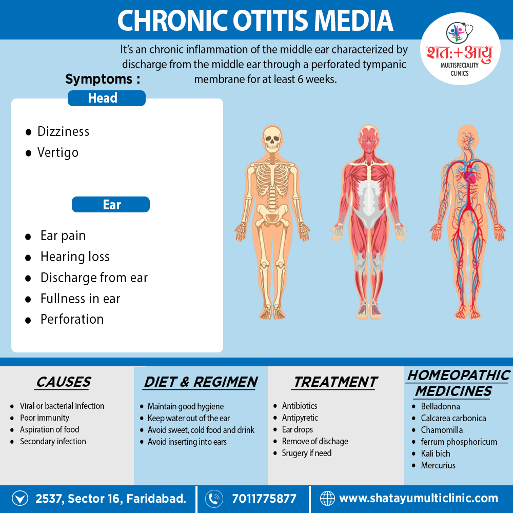
Chronic suppurative otitis media (CSOM) is a chronic inflammation of the middle ear and mastoid cavity that is characterize by discharge from the middle ear through a perforated tympanic membrane for at least 6 weeks.

Homeopathic Medicines used by Homeopathic Doctors in treatment of Chronic Otitis Media?

  • Belladonna
  • Chamomilla
  • Calcarea Carb
  • Hepar sulph
  • Sulphur
  • Capsicum
  • Ferrum Phos
  • Kali bich
  • Lycopodium
  • Mercurius Sol
  • Tellurium

What are the symptoms of Chronic Otitis Media?

  • Ear discharge
  • Hearing loss
  • Perforation
  • Middle ear mucosa- red, oedematous and swollen
  • Bleeding

What are the causes of Chronic Otitis Media?

  • Following exanthemata’s fever
  • Infection From tonsils, adenoids and infected sinuses
  • Persistent mucoid otorrhoea

References:

  1. https://en.wikipedia.org/wiki/Otitis_media
  2. Disease of EAR, NOSE, AND THROAT&HEAD AND NECK SURGERY 6TH EDITIONS BY P.L Dhingra, Shruti Dhingra.
  3. https://hpathy.com/homeopathy-papers/homeopathy-for-the-management-of-otitis-media/