Opening Hours : Mon to Sat - 9am to 9pm / Sun - Closed

Diabetes Mellitus

Definition:

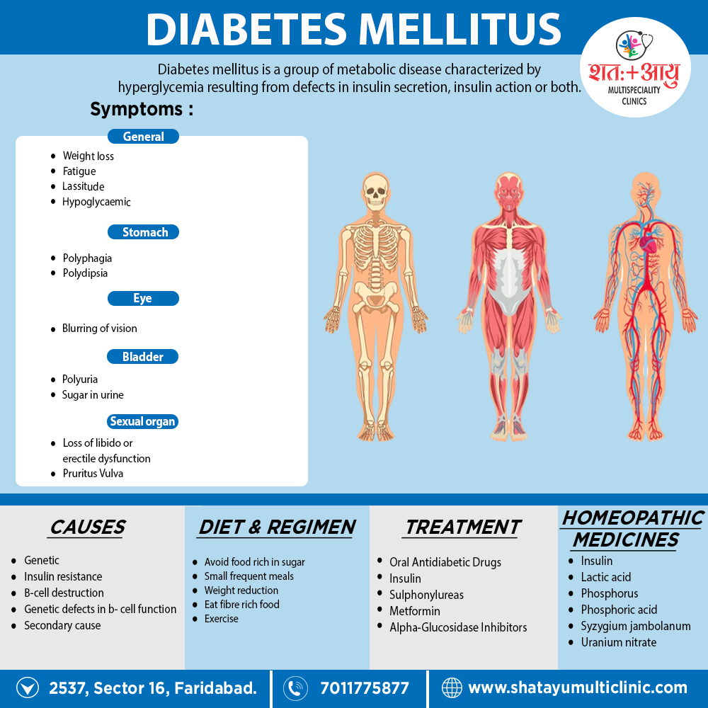
Diabetes mellitus is a group of metabolic diseases characterized by hyperglycaemia resulting from defects in insulin secretion, insulin action or both.[1]

Overview

Overview Diabetes Mellitus:

Diabetes mellitus is a disorder in which the amount of sugar in the blood is elevated. Doctors often use the full name diabetes mellitus, rather than diabetes alone, to distinguish this disorder from diabetes insipidus. Diabetes insipidus is a relatively rare disorder that does not affect blood glucose levels but, just like diabetes mellitus, also causes increased urination.[1]

Blood sugar

Generally, the three major nutrients that make up most food are carbohydrates, proteins, and fat. Sugars are one of three types of carbohydrates, along with starch also fiber.

Moreover, There are many types of sugar. Some sugars are simple, and others are complex. Table sugar (sucrose) is made of two simpler sugars called glucose and fructose. Additionally, Milk sugar (lactose) is made of glucose and a simple sugar called galactose. The carbohydrates in starches, such as bread, pasta, rice, and similar foods, are long chains of different simple sugar molecules. Sucrose, lactose, carbohydrates, also other complex sugars must be broken down into simple sugars by enzymes in the digestive tract before the body can absorb them.

Besides this, Once the body absorbs simple sugars, it usually converts them all into glucose, which is an important source of fuel for the body. Glucose is the sugar that is transported through the bloodstream and taken up by cells. All in all, The body can also make glucose from fats and proteins. Blood “sugar” really means blood glucose.[1]

 Insulin

Basically, Insulin, a hormone released from the pancreas (an organ behind the stomach that also produces digestive enzymes), controls the amount of glucose in the blood. In detail, Glucose in the bloodstream stimulates the pancreas to produce insulin. Insulin helps glucose to move from the blood into the cells. Besides this, Once inside the cells, glucose is converted to energy, which is used immediately, or the glucose is stored as fat or the starch glycogen until it is needed. Lastly, The levels of glucose in the blood vary normally throughout the day.

Level of Glucose

They rise after a meal and return to pre-meal levels within about 2 hours after eating. In detail, Once the levels of glucose in the blood return to pre-meal levels, insulin production decreases. The variation in blood glucose levels is usually within a narrow range, about 70 to 110 milligrams per decilitre (mg/dL), or 3.9 to 6.1 millimoles per liter (mmol/L) of blood in healthy people. If people eat a large amount of carbohydrates, the levels may increase more. People older than 65 years tend to have slightly higher levels, especially after eating.

If the body does not produce enough insulin to move the glucose into the cells, or if the cells stop responding normally to insulin (called insulin resistance), the resulting high levels of glucose in the blood also the inadequate amount of glucose in the cells together produce the symptoms and complications of diabetes.[1]

Classification

Classification of Diabetes Mellitus:

Etiological Classification of Diabetes mellitus and Impaired glucose tolerance

1.Type I DM (in other words; β-cell destruction, absolute insulin deficiency).

Insulin dependent diabetes mellitus (IDDM)

(a) Autoimmune

(b) Idiopathic

2.Type II DM – Non-insulin dependent (in other words; NIDDM)

3. Maturity onset diabetes in young (in other words; MODY 1-6)

Genetic defects in β-cell function have the following

mutations:

  • MODY 1 – Hepatocyte Nuclear Transcription factor (HNF) 4α
  • MODY 2 – Glucokinase
  • MODY 3 – HNF – 1α
  • MODY 4 – Insulin promotor factor-1 (IPF1)
  • MODY 5 – INF – 1β
  • MODY 6 – Neurogenic differentiation1, (Neuro D1)

4.Secondary causes

a. Pancreatic disease i.e.
  • Cystic fibrosis
  • Trauma / pancreatopathy
  • Hemochromatosis
  • Pancreatitis
  • Fibrocalcific pancreatic diabetes (in other words; FCPD)
  • Carcinoma of pancreas
b. Endocrine disorder i.e.
  • Acromegaly
  • Cushing syndrome
  • Glucagonoma
  • Conn’s syndrome
  • Pheochromocytoma
  • Hyperthyroidism
c. Drug induced i.e.
  • Glucocorticoids
  • Diazoxide
  • Thiazides
  • Phenytoin
  • Pentamidine
  • Alpha Interferon
  • Thyroid hormone
  • Beta adrenergic
d. Infection i.e.
  • Congenital Rubella
  • Coxsackie B virus
  • Cytomegalovirus
e. Insulin Receptor Defect i.e.
  • Anti-insulin Receptor Antibodies [in other words; Stiffman syndrome]
  • Lipoatrophic Diabetes
f. Other Genetic syndrome associated with DM i.e.
  • Down’s Syndrome
  • Turner’s Syndrome
  • Wolframe’s syndrome
  • Prader willi syndrome
  • Kline filters syndrome
  • Porphyria
  • Myotonic Dystrophy
  • Laurence-Moon-Biedl syndrome
  • Friedreich’s ataxia

5.Gestational diabetes

6.Impaired glucose tolerance (Borderline diabetes)

a. Primary: Obese, non-obese

b. Secondary: All conditions mentioned under secondary DM, cirrhosis of liver, kidney failure, chronic undernutrition, hypokalaemia, stress, e. g. myocardial infarction.[1]

Pathogenesis

Pathogenesis of Diabetes Mellitus:

Type 1 diabetes i.e.

Due to a combination of genetic, autoimmune and environmental factors, there is destruction of pancreatic β cells.

Genetic factors i.e.–

(a) Twin studies: Among monozygotic twins, the concordance rate is 30–70%, i.e., if one twin gets DM, the chances of second twin getting are 30–70%. (b) Major Histocompatibility Complex (MHC). Genes on the short arm of chromosome 6 Encodes for Human Leucocyte Antigens (in other words; HLA). There Is a strong association with Type I DM and HLA-DRD and HLA-DR4.

Autoimmunity

leads to lymphocytic infiltration of the islets of Langerhans called insulinitis. β cells are more susceptible to destruction by cytokines like TNF-α, TNF-γ IL-1.

Autoimmune basis is also suggested by association with other autoimmune diseases like Graves’ disease, Coeliac disease, pernicious anaemia. Addison’s disease, polyglandular autoimmune syndrome I and II.

Environmental Factor

(a) Viruses: Congenital rubella, Rota virus, mumps

(b) Chemical: Nitrosourea compounds, Rodenticide,

Pentamidine are associated with increased risk.

(c) Diet: (i) Cow’s milk (bovine milk proteins) may

stimulate immune response against β cells. (ii)

Type 2 Diabetes (NIDDM)

Etiopathogeneses

Type 2 DM is disorder characterized by insulin resistance and impaired insulin secretion. Hyperglycemia develops due to (i) Peripheral resistance to action of insulin. (ii) Increased hepatic glucose output. (iii) Impaired pancreatic β- cell secretion of insulin.[1]

        1.Genetic factors:

Type 2 DM has very strong genetic predisposition.

(a) Firstly, Twin studies – Concordance in monozygotic twins is 70–90%. If both parents are diabetic, their off spring has 90% chance of developing type 2 DM.

(b) Secondly, Polygenic factors – Type 2 DM is a polygenic multifactorial disease, in majority of cases. No consistent major genetic genes have yet been implicated.

(c) Thirdly, Monogenic forms are rare, e.g., Wolfram syndrome, MODY types 1-6, MELAS (e.g. mitochondrial myopathy, encephalopathy, lactic acidosis, stroke).

2.Obesity lipotoxicity, nutrition i.e.

(a) Obesity and central fat distribution. The cut offs for BMI are 23 kg/m2 and waist circumference for men at 90 cm and for women 80 cm.

(b) Lipotoxicity. Adipose tissue can secrete a large number of ‘adipokines’ which can impair insulin secretion and action by an endocrine effect, e.g. leptin or increase in insulin resistance, e.g., TNFα,

Resist in, Adiponectin (low levels contribute to Insulin resistance). Free fatty acids inhibit glucose uptake and increase hepatic glucose output

3.Nutrition i.e.:

Generous dietary intake Physical inactivity Vitamins and trace elements – Low levels of Vitamin D, zinc, copper, chromium.

4.Insulin resistance i.e.

It is a state in which a given concentration of insulin gives a subnormal biological response on patients target tissue e.g., skeletal muscle, adipose tissue and liver. There is resistance to exogenous and endogenous insulin. There are overt diabetics who do not respond to conventional and supraphysiological levels of insulin, but at the end are commonly seen in individuals with mild glucose intolerance. It is also called metabolic syndrome. Deadly quartet-CHAOS (CAD, HT, Obesity, Adult-Onset diabetes and Stroke).

5.The ‘thrifty phenotype’ hypothesis i.e.

Individuals with low birth weight appear to have higher risk of NIDDM, particularly if they become obese in later life. This hypothesis suggests that intra uterine malnutrition leads to defective pancreatic development. Such individuals may become susceptible to diabetes, hypertension and heart disease in later life.[1]

6.Criteria for diagnosis of IR (WHO)
  • Central obesity (BML, WHR)
  • Low Tg (>150 mg/L, high LDL)
  • Low HDL (< 40-50 mg/dL)
  • High BP (>130/85 mm Hg)
  • FBS >100 mg/dL

Risk factors for type 2 diabetes

  • Age ± 45 years
  • Overweight (BMI ≥ 25 kg/m2)
  • Family history of diabetes (e.g. parents or siblings with diabetes)
  • Habitual physical inactivity
  • Previously identified IFG or IGT
  • History of GDM or deliver of a baby >9 lbs
  • Hypertension (≥ 140/90 mm Hg in adults)
  • HDL cholesterol ≥35 mg/dL (0.9 mmol/L) and/or triglyceride level ≥250 mg/dL (2.82 mmol/L)
  • Polycystic ovary syndrome
  • History of vascular disease

Wheat proteins (for example; gluten). Gluten-sensitive enteropathy (in other words; coeliac disease) in 5–10% cases.

(d) Stress in early life

Sign & Symptoms

Symptoms and Sign of Diabetes Mellitus

Onset –

Usually gradual in adults, but acute in children.

Modes of presentation –

Presence of osmotic symptoms (i.e. polyuria, polyphagia, polydipsia)

  • Weight loss, fatigue and lassitude
  • Pruritus vulvae in females or balanitis in males
  • Loss of libido or erectile dysfunction
  • Blurring of vision
  • Symptoms due to diabetes – related complications e.g., abdominal pain in ketoacidosis [1]

Convulsions – Fits should be considered hypoglycaemic until proved otherwise, especially in children. EEG abnormalities are common in insulin-treated patients, especially those with recurrent hypoglycaemia. Day Time attacks suggest epilepsy, while nocturnal attacks are often hypoglycaemic.

Investigation

Blood glucose estimation

In non-diabetes, blood glucose levels are 70-100 mg/dl.

(a) Firstly, Random blood glucose is done at any time in the day, irrespective of meals. If <140 mg normal, >200 mg indicates DM and between 140–200 mg, GTT is required.

(b) Secondly, Fasting blood glucose is measured after 12 hr fast. Normal <100 mg, diabetes if >126 mg and impaired fasting glucose 101–125 mg (pre-diabetics).

(c) Thirdly, Post-prandial blood glucose is measured 2 hr after a meal. Normal <140 mg, Diabetes >200 mg Impaired glucose tolerance 140–199 mg.

Oral Glucose tolerance test (in other words; OGTT)

Pre diabetes also known as impaired glucose tolerance is a condition with no symptoms. But this patient develops more serious DM. Hence individuals with family history DM 2, Women with gestational diabetes or had a baby weighing >9 pounds, the obese with much belly fat and elderly should go under the test. In OGTT the first sample taken overnight fast of 8 hrs following which patient is given glucose 1 g/kg body wt. Blood is then collected at 30, 60, 120, 180 minutes. In diminished Glucose tolerance, a level of blood glucose is raised at 180 minutes.

Factor affecting GTT i.e.
  1. Age – GTT decreases in patient > 60 years of age
  2. Obesity
  3. Surgery – Due to stress of surgery, there is increase in cortisol and catecholamine secretion.
  1. Drugs – For example; Thiazides, phenytoin, oral contraceptives, thyroxine, corticosteroids.
Urine glucose

In brief, Normally about 100 mg/d is excreted and can be estimated by tests for reducing sugar e.g. Benedict’s tests or clinistix method. Those measurements are now not recommended.

3.Glycosylated haemoglobin (GHb)

In general, It is used to measure long-term glycaemic control. Hb in the blood is glycated by sugar. In detail, Glucose combined with protein residues on Hb chain by slow ‘Non-enzymatic’ Glycation. This results in formation of Haemoglobin A1c proportionate to amount of blood glucose. Since life span of RBCs is 21 days, HbA1c will reflects blood glucose level over past 2-3 months.

Advantages – No relation to meals, physical activity, crash diets or modes of therapy.

Disadvantages –

(a) Firstly; Cannot be used to change daily diseases of drugs or insulin.

(b) Secondly; Values are unreliable in haemolytic anaemia and haemoglobinopathies because of reduced life span of RBCs.

(c) Thirdly; In iron deficiency anaemia, values are higher, lower values in pregnancy, Vitamin C, E deficiency.

Self-monitoring of Blood Glucose

  • Type I diabetes 4–6 times a day depending on the insulin regimens.
  • Type II diabetics on oral hypoglycaemic drugs need to monitor 1-2 times/d
  • During pregnancy more frequent monitoring.
  • During acute complications or illness 2-hourly.

Microalbumin. Pts with microalbuminuria have a greater risk of developing kidney failure, also cardiovascular damage. Furthermore; Reference is 0-1.7 mg/dl in spot random sample, up to 20 mg/L in 24 hr urine volume.

Urine protein/creatinine ratio gives information about proteinuria in pts with diabetic nephropathy.

Higher the ratio, greater the damage.

Serum creatinine.

In diabetics with nephropathy a doubling of creatinine suggests a 50% reduction in GFR. Presence of the antibody – (a) Is a strong predictive marker for onset of type 1 DM. (b) Helps differentiate between Type 1 and Type 2 diabetes. (c) Aids in prediction, diagnosis also management of DM. (d) Indicates need for progression to insulin therapy in Type 2DM. (e) To determine the cause of hypoglycaemia. (f) To monitor recovery after removal of an insulin producing tumour of the pancreas. Early detection of IDDM – Some of the markers that we can predict the development are given in.

Insulin levels.

Elevated blood glucose levels with low insulin levels indicate insufficient insulin for adequate control of blood glucose. Similarly, high insulin levels with low blood glucose indicate a change in dosage of drugs.

C-peptide

It is used to monitor beta cell activity and capability over time and help in deciding when to start insulin therapy. C-peptide in conjunction with insulin and glucose levels can be used to diagnose cause of hypoglycaemia and monitor its treatment.

Insulin antibody test

Quantitative determination of antibodies against insulin in serum.

GAD-65 antibody

It is an enzyme produced primarily by pancreatic islet cells. GAD-65 antibodies are common in newly diagnosed diabetic patients also often appear years before clinical onset of the disease.

Presence of the antibody – (a) Is a strong predictive marker for onset of type 1 DM. (b) Helps differentiate between Type 1 and Type 2 diabetes. (c) Aids in prediction, diagnosis and management of DM. (d) Indicates need for progression to insulin therapy in Type 2 DM. (e) To determine the cause of hypoglycaemia. (f) To monitor recovery after removal of an insulin producing tumour of the pancreas.

Complications

  • Metabolic Complications
  • Diabetic Ketoacidosis.
  • Hyperosmolar Hyperglycaemic State
  • Hypoglycaemic Coma
  • Diabetic Retinopathy
  • Diabetic Nephropathy
  • Pregnancy also Proteinuria in Diabetes
  • Diabetic Foot

In Pregnancy

Pregnancy and diabetes

Pregnancy predisposes to diabetes by three mechanisms.

  1. Antagonization of peripheral actions of insulin due

to raised levels of oestrogens, progesterone, corticosteroids, human chorionic somatomammotropin and human placental lactogen.

  1. Rapid insulin destruction due to high insulins activity of placenta.
  2. Depletion of β cell insulin reserve. Increased utilization of stored nutrients and glucose to feed the foetus leads to fasting hypoglycaemia, ketonemia and elevated free acids which leads to insulin release and ultimately depletion of beta cell insulin reserve.

Management of diabetes in pregnancy

  1. Diet high protein intake. Restriction of salt if oedema.
  2. Exercise
  3. Insulin therapy is essential. In first half of pregnancy requirement may be reduced due to morning sickness. Constant glucose utilization through the placenta and absence of antagonists to maternal glucose utilization. Post-partum insulin dosage must be readjusted as insulin requirements fall due to removal of placental lactogen.
  4. Oral hypoglycaemic agents should be avoided except metformin which is effective in women with PCOD to aid conception.[1]

Foetal monitoring in GDM for foetal anomalies

(a) USG at 18–22 weeks for malformations.

(b) Foetal echo at 20–24 wks.

(c) Serum α-fetoproteins.

(d) Chromosomal studies if required.

For foetal well-being

(a) Maternal records of foetal movements. (b) Foetal heart rate patterns (cardiotocography). (c) USG. Non-stress test, contraction. (d) Lecithin: Sphingomyelin ratio (for lungs). Renal glycosuria is a rare condition in which sugar is eliminated in urine despite a normal or low blood sugar.

This is due to improper functioning of renal tubules. When glucose in blood increases beyond 180 mg/dL, it is excreted in urine. This point is known as renal threshold for glucose (RTG).

Adverse effects of diabetes on pregnancy

  • Polyhydramnios
  • Pre-eclampsia, eclampsia
  • Recurrent abortions
  • Premature labour
  • Prolonged labour
  • Abnormal presentations
  • UTI
  • Monilial vaginitis
  • Hypoglycaemia (first trimester)
  • Hyperglycaemia (third trimester)
  • Postpartum haemorrhage
  • Puerperal sepsis
  • Retinopathy
  • Nephropathy
  • Gastropathy

Adverse effect on foetus

  • Macrosomia (large babies) because of foetus/hyper-insulinemic causing excessive fat deposition and visceromegaly.
  • Prematurity.
  • Post-partum hypoglycaemia from persisting insulin secretion after birth in absence of glucose supplied via placenta.

• Intra-uterine death more common after 37th week, hence early termination of pregnancy is advisable especially if she has vascular complications.[1]

Special Category

  1. Late-onset Autoimmune Diabetes of Adults (LADA) –A variation of type 2 DM, patients have autoanti bodies such as anti-islet cell and anti-glutamic acid (GAD) antibodies. Insulin levels are low. However, they progress gradually and present in non-obese young adults and may respond to OHAs. Eventually they became insulin dependent.
  2. Maturity onset Diabetes of the Young (MODY2) is

inherited genetic defect in β-cell glucokinase function inherited genetic defect in β-cell glucokinase function defect. Patients are detected during routine screening or during pregnancy. The hyperglycaemia is minimal. MODY 1,3,4,5,6 present in early adulthood, are symptomatic, have severe hyperglycaemia and are prone to develop complications.

  1. Fibro Calculous Pancreatic Diabetes (FCPD). See later.

4. Brittle diabetes – Here patients have wide fluctuations in blood sugar and recurrent episodes of ketoacidosis in spite of adequate doses of insulin. It usually occurs in young females with psychiatric problems and menstrual irregularities.[1]

Diabetic Ketoacidosis

Diabetic Ketoacidosis

Is a state of acidaemia induced by excess production of ketoacids, Dehydration and hyperglycaemia are the rule, lactic acidosis may also be present.[1]

Pathophysiology

Diabetic ketoacidosis is caused by severe insulin deficiency and is accentuated by excessive glucagon secretion. This leads to major clinical and laboratory abnormalities seen in diabetic ketoacidosis, which includes excess mobilization of free acids from adipose tissue, increased glucose production from the liver and impaired glucose uptake and utilization by muscle.

The two major effects of uncontrolled diabetes are:

  • Increased glucose production which causes hyperglycaemia, osmotic diuresis, electrolyte depletion and dehydration
  • Increased ketogenesis, resulting in metabolic acidosis.

Diagnosis

The cardinal features are:

  • Acidosis (arterial pH ≤ 7.3)
  • Plasma anion gap (≥ 16 mmol/liter)
  • Serum ketone is positive
  • Serum bicarbonate ≥ 15 mmol/liter
  • Hyperglycaemia (plasma glucose ≥ 11.1 mmol/liter)

Investigations:

Blood glucose, urea and electrolytes (especially potassium), full blood count and blood gases. ECG should be monitored continuously for signs of hypokalaemia.[1]

Management:

Admission

  • Diagnosis suspected and confirmed immediately by blood glucose and ketone measurements
  • Initial assessment of magnitude of dehydration, hyperosmolality, and acidosis
  • Fluid loss measured by subtracting admission weight from last recently known stable weight
  • Effective serum osmolality = 2 × [serum Na+ (mmol/liter) + serum K + (mmol/liter)] + serum glucose (mmol/liter) + urea
  • Evaluate patient for sepsis and/or precipitating illness [1]
Hour 1
  • Fluid administration

– If strikingly hypovolaemic with low blood pressure and relative or absolute anuria, fluid administration should be normal saline and, if necessary, colloids; rate of administration should be that necessary to restore circulatory function

– When blood pressure is normal and urine output adequate: fluid administration should be normal saline; rate of (mmol/liter)] administration 1000 mL/hour

  • Initial assessment of serum potassium and kidney function.
  • Insulin

– Continuous intravenous infusion of regular insulin 5–10 units/ hour or intramuscular regular insulin (20 units loading dose and 5 units/hour) Potassium

– Start intravenous potassium at 10–40 mmol/hour at initiation of insulin therapy if serum potassium is not > 5 mmol/litter and renal output is good. If patient is hyperkaliaemic, temporary delay intravenous potassium Alkali

– Sodium bicarbonate intravenously is seldom indicated, except in severe acidosis (pH < 7.0) with incipient circulatory collapse Dose, if given, is 50–100 mmol/litter sodium bicarbonate given in 0.45% saline over 30–60 minutes. Additional K + must be given with bicarbonate therapy

Hour 2
  • Fluid administration
  • Continue normal saline at 500 mL/hour. Maintain calculated plasma osmolality > 285 mOsmol/liter throughout the first 12 hours. If serum Na + >150 mmol/liter switch to 0.45% saline
  • Insulin
  • Check blood glucose and adjust insulin dose to maintain a fall of about 5 mmol/litre/hour. Do not allow blood glucose to fall below 11.1–14.0 mmol/litre. Anion gap should be decreasing and blood pH increasing
  • Potassium
  • Maintain serum potassium at 4.0–5.0 mmol/litre by continued addition of potassium to intravenous fluids[1]
Hours 3-4

Continue as for hour 2

Observe for cognitive or neurological symptoms and continue to do so for 12 hours

Hours 5-8
  • Fluid administration

– Normal saline 250 mL/hour. When blood glucose reaches 11.1–14.0 mmol/liter, change intravenous fluid to 500 mL/ hour normal saline with 5% glucose

  • Insulin

– Continue insulin at maintenance dose until ketoacidosis has cleared (blood pH > 7.35, serum ketones negative)

  • Potassium

– Continue at 10–40 mmol/hour until ketoacidosis has cleared

  • Phosphate

– Consider phosphate replacement at 6 hours if serum phosphate is < 2.0 mg/dL

Hours 8-24
  • Fluid administration

– Continue intravenous repletion with 0.45% saline with or without 2.5% or 5.0% glucose as needed

  • Insulin

– After ketoacidosis has cleared (blood pH > 7.35, serum ketones negative) switch to subcutaneous insulin and then stop intravenous or intramuscular insulin.

Complications of DKA

Iatrogenic complications
Osmotic and volume disturbances–

Administration of insulin without sufficient fluids causes shift of water from extracellular space; further shrinking of extracellular fluid volume impairs blood flow to critical vascular beds, or precipitates vascular collapse. Similarly, insufficient saline administration may also result in hypotension.

Potassium disturbances –

Premature (before insulin has begun to act) and inappropriate potassium administration may cause fatal hyperkalaemia (cardiotoxicity) in early course of management. Glucose, insulin, and volume expansion with normal saline are potential modalities for lowering serum potassium; hence failure to administer potassium in latter stages may cause fatal hypokalaemia in potassium depleted patient.[1]

Hypoglycaemia and reappearance of ketosis –

During therapy of DKA, normalization of blood glucose usually achieve sooner than reversal of ketoacidosis state. Because insulin therapy must continue, hypoglycaemia develops unless glucose is given.

Fingerstick glucose measurements should done hourly for 4h, 2 hourlies for next 4h, and 4 hourlies till pt. improves. Glucose generally falls at rate of 50–100 mg/ dL/h. Failure to maintain glucose and insulin treatment until ketones have clear and depleted glycogen stores restored, results in recurrence of ketosis

Cerebral oedema –

It may rarely occur in children but is even rarer in adults. The condition should suspect when a pt. with ketoacidosis begins to deteriorate 3 to 10 hours into treatment with increasing stupor or coma coupled with signs of raised intracranial pressure; an unexpected fever may also an early sign. Osmotic disequilibrium between intracellular and extracellular fluids probably plays a role. Tr. – Mannitol 20% iv 1.5–2 mL/kg body wt. over 15 minutes and dexamethasone 10 mg iv followed by 4mg in q6h. Clinical response will seen in 12–24 h.

Hypocalcaemia –

It may develop during phosphate replacement.[1]

Non-iatrogenic complications

Infection –

Although leucocytosis may occur in DKA in absence of infection, fever generally indicates infection and demands careful search for pneumonia, pyelonephritis and septicaemia. A Rare ketoacidosis-associated infection is mucormycosis of paranasal sinuses with facial pain, bloody nasal discharge, orbital swelling, blurred vision and impaired consciousness. Tr. – Broad spectrum antibiotics. Blood, Urine and throat cultures should be taken prior to giving antibiotic.

Vascular thrombosis –

Combination of volume contraction, low cardiac output, increased blood viscosity, underlying atherosclerosis, direct endothelial damage due to hyperosmolar milieu, and changes in clotting factors and platelet function predispose to thrombosis.

Respiratory distress syndrome i.e.–

Hypoxia and ARDS may develop in course of therapy of DKA hyperosmolar coma. Additionally, Clinical picture is characterized by unexplained hypoxaemia and dyspnoea in absence of any underlying pulmonary/cardiac disease.

Pancreatitis i.e.–

Acute abdominal pain is a common presentation of DKA, and resolves rapidly on therapy. specific aetiologies can be gastroparesis, ischaemic bowel, and cholecystitis. Raised serum amylase is observed in 80% of cases. It may represent pancreatic damage (in other words; hypertonicity and hyper perfusion); in some instances, it is subclinical.

Myocardial infarction i.e.–

can precipitate or complicate DKA, it is a major cause of morbidity.

Hyperlipidaemia –

Severe hypertriglyceridemia (triglycerides > 1000 mg/dl) is seen in about 10% which on follow-up resolve in 70% of cases. The abnormality is related to acute metabolic changes in DKA.[1]

Treatment

Total calories requirements are determined by the patient’s activity

Overweight NIDDM should ne encourage to establish within their weight within a desirable range. A reduction of approximately 500 kcal/day can result in loss of 1–2 kg/month.

Carbohydrates i.e.

should comprise 55–60% of the calories, With the form and amount to determine by individual eating habits also blood glucose and lipid responses. Unrefined carbohydrates should substitute by refined carbohydrates to the extent possible

Proteins i.e.

In brief, Recommended dietary allowance of 0.85 g/kg body weight for adult is an appropriate guide. If kidney dysfunction, reduce intake to 0.6 g/kg.

Fat i.e.–

should comprise ≤ 30% of total calories and all components should reduce proportionately. Replacement of saturated with polyunsaturated fat is desirable to reduce cardiovascular risk. Cholesterol intake should < 300 mg/day.

Fibre i.e.

Increased consumption of dietary fibre especially soluble fibre associate with lower levels of blood glucose and serum lipids. The Water insoluble fibres such as cellulose, lignin and most hemicelluloses found in whole grain breads, cereals and wheat bran affect gastrointestinal transit time and faecal bulk with little impact on plasma glucose. However highly viscous water-soluble fibres such as pectins, gums and storage polysaccharide found in fruits, legumes, lentils roots, tubers, oat and oatbran, when eaten in purified form, reduce serum levels of glucose and insulin. Ideal recommended amount of fibre in patient’s diet is 35–40 g/day.

Alternative sweeteners i.e.

Both nutritive and non-nutritive sweeteners are acceptable in diabetes management.[1]

Sodium

Should restrict to 1000 mg/1000 kcal, not to exceed 3000 mg/d to minimize symptoms of hypertension.

Alcohol

in moderation and may need to restrict entirely by person with diabetes and insulin-induced hypoglycaemia, neuropathy, poor control of glucose and lipids, or obesity. Vitamins, minerals and antioxidants – intake should encourage.

  • Forbidden foods – e.g. Sugar, jam, jellies, honey, jaggery, tinned fruits and juices, sweets, chocolate, ice creams, pastries, glucose drinks, foods made with sugar, pudding, sauces.

(b) Foods allowed in moderation – e.g. Bread of all kinds and chapattis made from wheat or millets, plain biscuits, all fresh fruits, baked beans, breakfast cereals.

(c) Free foods – e.g. All meat, fish, eggs (not fried), clear soup or meat extracts; tea or coffee; vegetables such as cabbage, cauliflower, spinach, pumpkin, brinjal, lady’s finger, turnip, French beans, cucumber, lettuce, tomato, spring onions, radish, asparagus. Spices, salt, pepper and mustard; butter and margarine. Sugar Substitutes For sweetening.

Exercise

Medical evaluation prior to formulating exercise programme, History and physical examination i.e.

  • Review of diet and medication
  • Fundoscopy
  • Foot evaluation

Neurological evaluation –

(a) Firstly, Sensory/motor.

(b)  Secondly, Autonomic

  • Cardiovascular risk-factor profile
  • Stress test assessment of glucose control
Exercise prescription

Type must adjust to patient’s preference and existing medical condition. Aerobic exercise is preferred (e.g. swimming, cycling, walking, running). Addition of moderate resistance should be considered.

Duration – 20–45 mins. per session

Frequency 3-4 sessions per week is required to observe beneficial metabolic effect. 4-5 session per week for weight reduction.

Risk and benefits of exercise
Potential benefits of exercises i.e.
  • Maintenance of desirable body weight
  • Improved sense of wellbeing also enhanced social interactions
  • Enhanced insulin sensitivity
  • Improved glucose control
  • Decreases triglycerides also increases HDL-cholesterol levels
  • Improved fibrinolysis
  • Improved cardiac performance
Potential risks of exercises
  • Cardiovascular – Either, Myocardial ischaemia or infarction, dysrhythmias. Hypertensive response to exercise.
  • Microvascular – For Example; Retinal haemorrhage, proteinuria, accelerated microvascular disease.
  • Metabolic – For Example; Hypoglycaemia, hyperglycaemia, ketosis.
  • General – For Example; Muscle strains/sprains, foot injury, joint injury, degenerative arthritis, poor dietary compliance.

ADA recommends 150 min/week (distributed over at least 3 days) of moderate aerobic physical activity with no gaps longer than 2 days.

Intensity – Patient’s maximal pulse rate estimate by subtracting patient’s age from 220. Patient can start at 40 to 50% of their maximal pulse rate with gradual increments to 60–70% over 6–8 weeks.

Programme

  • Stretching (5–10 mins)
  • Warm up (5–10 mins)
  • Exercise (20–45 mins)
  • Warm down (10 mins at 30% of full exercise intensity) Exercise should be avoided when plasma glucose concentration is > 250 to 300 mg/dL and/or presence of ketones in urine.[1]

Oral Antidiabetic Drugs (OADs)

[1] Insulin Secretagogues.

Sulphonylureas

[2] Insulin Sensitizers

Biguanides: Metformin

[3] Thiazolidinediones

[4] Alpha-glucosidase Inhibitors

[5] Sodium-Glucose Co-Transporter 2 Inhibitors (in other words; SLGT2)

[6] Insulin

Homeopathic Treatment

Homeopathic Management

Homeopathic treatment is based on individualization in which a doctor selects a medicine according to your/ patients constitution rather than matching only symptoms similarity, so before taking any homoeopathic medicine you have to firstly consult a homoeopathic physician for your concern problem’s, there are so many remedies are useful for this condition but some few therapeutic indications of homeopathic remedies in the cases of Diabetes mellitus are as below.


Uranium nitrate. [Uran]
This remedy is praised highly by Hughes also others in diabetes originating in dyspepsia. It has polyuria, polydypsia, dryness of the mouth and skin. Additionally, It causes sugar in the urine. Dr. Laning said that no remedy gives such universally good results; it lessens the sugar and quantity of the urine; he recommended the 3X trituration. Besides this; It is when the disease is due to assimilative derangements that Uranium is the remedy, and symptoms e.g. defective digestion, languor, debility and much sugar in the urine, enormous appetite and thirst, yet the patient continues to emaciate.[2]


Syzygium jambolanum

It is a remedy capable of diminishing the amount of sugar in the urine, especially when used in the tincture also lower trituration, and some cases have reported cured, but it cannot consider a reliable drug, and its use seems entirely palliative. [2]

Phosphoric acid [Phos]

corresponds to diabetes of nervous origin; the urine is increased, perhaps milky in color and containing much sugar. In detail; It suits cases due to grief, worriment and anxiety, those who are indifferent and apathetic, poor in mental and physical force. It is unquestionably curative of diabetes mellitus in the early stages, great debility and bruised feeling in the muscles. Moreover, There will be loss of appetite, sometimes unquenchable thirst and perhaps the patient will be troubled with boils.

Besides this, When patients pass large quantities of pale colorless urine or where there is much phosphatic deposit in the urine it is the remedy. It thus may be a remedy in the form known as diabetes insipidus. Hering considered Plumbum one of the most important remedies in diabetes mellitus. Causticum, Scilla also Strophanthus may be of use in diabetes insipidus. All in all, Lycopodium cured a case in a weary, wretched patient; emaciated, increased appetite also great thirst; pale, profuse urine. Eight quarts in 24 hours.

Phosphorus. [Phos]

Useful in diabetes also pancreatic diseases, especially in those of a either tuberculous or gouty diathesis. The pancreatic involvement will call attention to Phosphorus. Natrum sulphuricum corresponds to the hydrogenoid constitution, with dry mouth also throat, and Arsenicum should be studied in diabetic gangrene, thirst and emaciation. Besides this; Sudden and extreme dryness of the mouth and marked physical restlessness are also guiding symptoms to this remedy, especially with a dark watery stool. Dr. P.Jousset reports positive success where the mouth is dry; frequent, abundant urination and tendency to skin eruption.[2]

Lactic acid. [Lact]

Generally, An exceedingly good remedy in the gastrohepatic variety of diabetes and good results often follow its use. It has a fine clinical record. Furthermore, The symptoms are: urinates copiously and freely, urine light yellow also saccharine, thirst, nausea, debility, voracious appetite also costive bowels. Dry skin, dry tongue, gastralgia. Acetic acid also a valuable diabetic remedy, and it has passing of large quantities of pale urine, intense thirst, hot, dry skin and marked debility Carbolic acid may also found useful.[2]

Bryonia. [Bry]

Should not neglected in this disease. In brief, No remedy has dryness of the lips as a symptom of hepatic disorder more marked than Bryonia, also this is often one of the first symptom of diabetes. There is a persistent bitter taste, the patient is languid, morose also dispirited, thirst may not extreme nor the appetite voracious, the patient may lose strength through inability to eat. Podophyllum has a bitter taste, but the tongue is flabby. It may of use in the disease.

Chionanthus a remedy use by the Eclectic School upon the indications of thirst, frequent also copious urine; constipation with stools light colored, devoid of bile. Functional liver disorders. Argentum metallicum. Hahnemann suggests the use of this remedy especially, in diuresis, it decide use in diabetes insipidus. The urine is profuse, turbid also of sweet odor. Micturition is frequent and copious. Natrum sulphuricum. Hinsdale reports good results with this remedy. It has polyuria, intense itching of the skin, especially upon the upper surface of the thighs. Lastly,  It is the Tissue Remedy of diabetes.

Insulin. [Insulin]

Long before the discovery of Insulin Dr.Pierre Jousset of Paris prepared a pancreatic juice on a glycerine basis which he administered to diabetic patients in doses of 10 or 20 drops a day in water also had results sufficiently good to consider pancreatic juice, orally administered, as a remedy of great value in diabetes. Dr. Cartier, his practical successor, praised it insisted on smaller doses given by mouth as larger doses and hypodermic injections of it had no effect in ordinary diabetes.

Baker advises the homoeopathic strengths of Insulin 3d to 30th and reports happy results therefrom. Additionally, Great care must be taken not to overdose. Boericke says that it maintains the blood sugar at a normal level and the urine remains free of sugar. Lastly,  Epileptic convulsions also mental derangements have been produced by hypodermic use of this hormone.[2]

FAQs

Frequently Asked Questions

What is Diabetes Mellitus?

Diabetes mellitus is a group of metabolic diseases characterized by hyperglycaemia resulting from defects in insulin secretion, insulin action or both.

What causes Diabetes Mellitus?

  • β-cell destruction, absolute insulin deficiency
  • Mutations: Glucokinase, HNF – 1α, Insulin promotor factor-1
  • Pancreatic disease
  • Endocrine disorder
  • Drug induced
  • Infection
  • Insulin Receptor Defect
  • Down’s Syndrome
  • Turner’s Syndrome

Homeopathic Medicines used by Homeopathic Doctors in treatment of Diabetes Mellitus?

  • Uranium nitrate
  • Syzygium jambolanum
  • Phosphoric acid
  • Phosphorus
  • Lactic acid
  • Bryonia

What are the symptoms of Diabetes Mellitus?

  • Polyuria, Polyphagia, also Polydipsia
  • Weight loss, fatigue and lassitude
  • Pruritus vulvae in females
  • Balanitis in males
  • Either Loss of libido or erectile dysfunction
  • Blurring of vision
  • Abdominal pain in ketoacidosis

What are the types of Diabetes Mellitus?

  • Type I DM
  • Type II DM
  • Maturity onset diabetes in young
  • Secondary causes
  • Gestational diabetes
  • Impaired glucose tolerance

References:

  1. Text book of medicine by Golwala
  2. Therapeutics from Zomeo ultimate LAN