Opening Hours : Mon to Sat - 9am to 9pm / Sun - Closed

Acute Otitis Media

Definition:

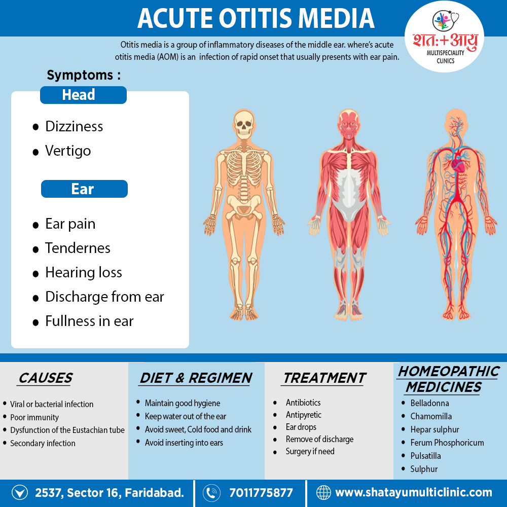
Otitis media is a group of inflammatory diseases of the middle ear. where’s acute otitis media [AOM] is an infection of rapid onset that usually presents with ear pain.[1]

Overview

Overview:

It is more common especially in infants and children of lower socioeconomic group. The cause of AOM is related to childhood anatomy and immune function. Typically, the disease follows viral infection of upper respiratory tract but soon the pyogenic organisms invade the middle ear.[1]

Causes

Cause:

The common cause of all forms of otitis media is

1. Dysfunction of the Eustachian tube.

This is usually due to inflammation of the mucous membranes in the nasopharynx, which can be caused by a viral upper respiratory tract infection (URTI), strep throat, or possibly by allergies.

2. By reflux or aspiration,

of unwanted secretions from the nasopharynx into the normally sterile middle-ear space, the fluid may then become infected — usually with bacteria.

3. upper respiratory infection:

The virus/bacteria that caused the initial upper respiratory infection can itself be identified as the pathogen causing the infection. Bacteriology. Most common organisms in infants and young   children are Streptococcus pneumoniae (30%), Haemophilus influenzae (20%) and Moraxella catarrhalis (12%). In addition, Other organisms include Streptococcus pyogenes, Staphylococcus aureus and sometimes Pseudomonas aeruginosa. In about 18–20%, no growth is seen. Many strains of H. influenzae and M. catarrhalis are β-lactamase producing.[1][4]

Risk Factors

Predisposing factors:

Anything that interferes with normal functioning of eustachian tube predisposes to middle ear infection. It could be:

1.Recurrent attacks of common cold, upper respiratory tract infections also exanthemata’s fevers like measles, diphtheria or whooping cough.

2.Infections of tonsils also adenoids.

3.Chronic rhinitis also sinusitis.

4.Nasal allergy.

5.Tumours of either nasopharynx, packing of nose or nasopharynx for epistaxis.

6.Cleft palate.[4]

Pathophysiology

Pathophysiology of Acute Otitis Media

Otitis media begins as an inflammatory process following a viral upper respiratory tract infection involving the mucosa of the nose, nasopharynx, middle ear mucosa, and Eustachian tubes. Due to the constricted anatomical space of the middle ear, the edema caused by the inflammatory process obstructs the narrowest part of the Eustachian tube leading to a decrease in ventilation.

This leads to a cascade of events resulting in an increase in negative pressure in the middle ear, increasing exudate from the inflamed mucosa, and build up of mucosal secretions, which allows for the colonization of bacterial and viral organisms in the middle ear. The growth of these microbes in the middle ear then leads to suppuration and eventually frank purulence in the middle ear space.

This is demonstrated clinically by a bulging or erythematous tympanic membrane and purulent middle ear fluid. This must be differentiated from chronic serous otitis media (CSOM), which presents with thick, amber-colored fluid in the middle ear space and a retracted tympanic membrane on otoscopic examination. Both will yield decreased TM mobility on tympanometry or pneumatic otoscopy.

Several risk factors can predispose children to develop acute otitis media. The most common risk factor is a preceding upper respiratory tract infection. Other risk factors include male gender, adenoid hypertrophy (obstructing), allergy, daycare attendance, environmental smoke exposure, pacifier use, immunodeficiency, gastroesophageal reflux, parental history of recurrent childhood OM, and other genetic predispositions.

Types

Types:

[1] Acute otitis media (AOM),

It is an infection of rapid onset that usually presents with ear pain. In young children this may result in pulling at the ear, increased crying, and poor sleep. Additionally, Decreased eating and a fever may also be present.

[2] Otitis media with effusion (OME),

It is typically not associated with symptoms, although occasionally a feeling of fullness is described; also it defined as the presence of non-infectious fluid in the middle ear for more than three months. It is characterized by insidious onset of hearing loss, sometime delayed and defective speech in children of school going age and mild earache.

[3] Acute Necrotizing otitis media:

It is a variety of acute suppurative otitis media, often seen in children suffering from measles, scarlet fever or influenza. Causative organism is β-hemolytic streptococcus. In addition, There is rapid destruction of whole of tympanic membrane with its annulus, mucosa of promontory, ossicular chain also even mastoid air cells. There is profuse otorrhea. In these cases, healing is followed by fibrosis or ingrowth of squamous epithelium from the meatus (secondary acquired cholesteatoma).

[4] Recurrent Acute otitis media:

Infants and children between the age of 6 months and 6 years may get recurrent episodes of acute otitis media. Such episodes may occur four to five times in a year. Usually, they occur after acute upper respiratory infection, the child being free of symptoms between the episodes. In addition, Recurrent middle infections may sometimes be superimposed upon an existing middle ear effusion. Sometimes, the underlying cause is recurrent sinusitis, velopharyngeal insufficiency, hypertrophy of adenoids, infected tonsils, allergy also immune deficiency. Feeding the babies in supine position without propping up the head may also cause the milk to enter the middle ear directly that can lead to middle ear infection.[1][4]

Sign and Symptoms

Sign and Symptoms of AOM:

The primary symptom of acute otitis media is like:

  • Ear pain
  • Fever
  • Reduced hearing during periods of illness
  • Tenderness on touch of the skin above the ear
  • Purulent discharge from the ears
  • Irritability and diarrhea (in infants)

Since an episode of otitis media is usually precipitated by an upper respiratory tract infection (URTI), there are often accompanying symptoms such as,

  • Cough and nasal discharge.
  • Feeling of fullness in the ear

Discharge from the ear can be caused by acute otitis media with perforation of the eardrum, chronic suppurative otitis media, either tympanostomy tube otorrhea, or acute otitis externa. Trauma, such as a basilar skull fracture, can also lead to cerebrospinal fluid otorrhea (discharge of CSF from the ear) due to cerebral spinal drainage from the brain and its covering (meninges).[1][4]

Clinical Examination

Clinical Examination of Acute Otitis Media

Otoscopic examination should be the first and most convenient way of examining the ear and will yield the diagnosis to the experienced eye. In AOM, the TM may be erythematous or normal, and there may be fluid in the middle ear space. In suppurative OM, there will be obvious purulent fluid visible and a bulging TM.

The external ear canal (EAC) may be somewhat edematous, though significant edema should alert the clinician to suspect otitis externa (outer ear infection, AOE), which may be treated differently. In the presence of EAC edema, it is paramount to visualize the TM to ensure it is intact.

If there is an intact TM and a painful, erythematous EAC, ototopical drops should be added to treat AOE. This can exist in conjunction with AOM or independent of it, so visualization of the middle ear is paramount. If there is a perforation of the TM, then the EAC edema can be assumed to be reactive, and ototopical medication should be used, but an agent approved for use in the middle ear, such as ofloxacin, must be used, as other agents can be ototoxic.[5]

Investigation

Investigation of Acute Otitis Media

Laboratory Studies

Laboratory evaluation is rarely necessary. A full sepsis workup in infants younger than 12 weeks with fever and no obvious source other than associated acute otitis media may be necessary. Laboratory studies may be needed to confirm or exclude possible related systemic or congenital diseases.

Imaging Studies

Imaging studies are not indicated unless intra-temporal or intracranial complications are a concern.

  • When an otitis media complication is suspected, computed tomography of the temporal bones may identify mastoiditis, epidural abscess, sigmoid sinus thrombophlebitis, meningitis, brain abscess, subdural abscess, ossicular disease, and cholesteatoma.
  • Magnetic resonance imaging may identify fluid collections, especially in the middle ear collections.

Tympanocentesis

It may be used to determine the presence of middle ear fluid, followed by culture to identify pathogens.

Tympanocentesis can improve diagnostic accuracy and guide treatment decisions but is reserved for extreme or refractory cases.

Other Tests

Tympanometry and acoustic reflectometry may also be used to evaluate for middle ear effusion.[5]

Diagnosis

Diagnosis of AOM:

As its typical symptoms overlap with other conditions, such as acute external otitis, symptoms alone are not sufficient to predict whether acute otitis media is present; it has to be complemented by

[A] Physical examination of the tympanic membrane

Examiners may use a pneumatic otoscope with a rubber bulb attached to assess the mobility of the tympanic membrane.

  • To confirm the diagnosis, middle-ear effusion and inflammation of the eardrum have to identify; signs of these are fullness, bulging, cloudiness also redness of the eardrum.
  • After that, it is important to attempt to differentiate between acute otitis media and otitis media with effusion (OME), as antibiotics are not recommending for OME. It has suggesting that bulging of the tympanic membrane is the best sign to differentiate AOM from OME, with a bulging of the membrane suggesting AOM rather than OME.
  • Viral otitis may result in blisters on the external side of the tympanic membrane, which call bullous myringitis (myringa being Latin for “eardrum”).

[B]Tympanometry

[C] Reflectometry or hearing test

In more severe cases, such as those with associated hearing loss or high fever

[D] Audiometry

[E] Tympanogram

[F] Temporal bone CT and MRI 

can be used to assess for associated complications, such as mastoid effusion, subperiosteal abscess formation, bony destruction, venous thrombosis or meningitis.[1][4]

Differential Diagnosis

Differential Diagnosis

The following conditions come under the differential diagnosis of otitis media i.e.

  • Cholesteatoma
  • Fever in the infant and toddler
  • Fever without a focus
  • Hearing impairment
  • Pediatric nasal polyps
  • Nasopharyngeal cancer
  • Otitis externa
  • Human parainfluenza viruses (HPIV) and other parainfluenza viruses
  • Passive smoking and lung disease
  • Pediatric allergic rhinitis
  • Pediatric bacterial meningitis
  • Pediatric gastroesophageal reflux
  • Pediatric Haemophilus influenzae infection
  • Pediatric HIV infection
  • Pediatric mastoiditis
  • Pediatric pneumococcal infections
  • Primary ciliary dyskinesia
  • Respiratory syncytial virus infection
  • Rhinovirus (RV) infection (common cold)
  • Teething[5]

Treatment

Treatment of AOM:

Most cases of otitis media pass within a few days, so there’s usually no need to see your GP.

However, see your GP if you or your child have symptoms showing no sign of improvement after two or three days

Antibacterial therapy

Most ear infections clear up within three to five days and don’t need any specific treatment. If necessary, either paracetamol or ibuprofen should be used to relieve pain and a high temperature. Make sure any painkillers you give to your child are appropriate for their age.

  1. Decongestant nasal drops.
  2. Oral nasal decongestants.
  3. Analgesics and antipyretics.
  4. Ear toilet.
  5. Dry local heat helps to relieve pain.
  6. It is incising the drum to evacuate pus and is indicated when
  • drum is bulging and there is acute pain,
  • there is an incomplete resolution despite antibiotics when drum remains full with persistent conductive deafness and
  • there is persistent effusion beyond 12 weeks.

All cases of acute suppurative otitis media should carefully follow till drum membrane returns to its normal appearance and conductive deafness disappears.[4]

Prevention

Prevention

Breast-feeding, using family or small-group day care for infants and toddlers and avoiding exposure to household tobacco smoke are the main preventive measures against acute otitis media (AOM).

It is also useful to immunize children who have recurrent otitis media with the influenza and the pneumococcal vaccines. Antibiotic prophylaxis is the most effective method to reduce the frequency of new episodes of otitis in children with recurrent AOM, but it should be used with caution.

Tympanostomy tube placement and/or adenoidectomy can be considered as options in some situations. Now, probably, the best initial steps to take to prevent new episodes of otitis in children with recurrent AOM are antimicrobial treatment of each individual AOM episode and, in certain cases, antibiotic prophylaxis for short periods when an upper respiratory tract infection is present.[6]

Homeopathic Treatment

Homeopathic Treatment of Acute Otitis Media

Homeopathy treats the person as a whole. It means that homeopathic treatment focuses on the patient as a person, as well as his pathological condition. The homeopathic medicines selected after a full individualizing examination and case-analysis.

Which includes

  • The medical history of the patient,
  • Physical and mental constitution,
  • Family history,
  • Presenting symptoms,
  • Underlying pathology,
  • Possible causative factors etc.

A miasmatic tendency (predisposition/susceptibility) also often taken into account for the treatment of chronic conditions.

What Homoeopathic doctors do?

A homeopathy doctor tries to treat more than just the presenting symptoms. The focus is usually on what caused the disease condition? Why ‘this patient’ is sick ‘this way’?

The disease diagnosis is important but in homeopathy, the cause of disease not just probed to the level of bacteria and viruses. Other factors like mental, emotional and physical stress that could predispose a person to illness also looked for. Now a days, even modern medicine also considers a large number of diseases as psychosomatic. The correct homeopathy remedy tries to correct this disease predisposition.

The focus is not on curing the disease but to cure the person who is sick, to restore the health. If a disease pathology not very advanced, homeopathy remedies do give a hope for cure but even in incurable cases, the quality of life can greatly improve with homeopathic medicines.

Homeopathic Medicines for Acute Otitis Media:

The homeopathic remedies (medicines) given below indicate the therapeutic affinity but this is not a complete and definite guide to the homeopathy treatment of this condition. The symptoms listed against each homeopathic remedy may not be directly related to this disease because in homeopathy general symptoms and constitutional indications also taken into account for selecting a remedy, potency and repetition of dose by Homeopathic doctor.

So, here we describe homeopathic medicine only for reference and education purpose. Do not take medicines without consulting registered homeopathic doctor (BHMS or M.D. Homeopath).

Aconite [Acon]

Bayes recommends Aconite IX in the maddening pains of otitis, claiming it to be far superior to Chamomilla or Pulsatilla. Moreover, There is dark redness of the parts, stinging, lancinating or throbbing pains and great sensitiveness. It suits earache from sudden change of temperature; it is worse at night and aggravate by warmth. Its influence is restricting to a brief period immediately following exposure. In this respect Copeland says: “It differs from Ferrum phosphoricum, which has a much longer period of usefulness.”

Belladonna [Bell]

The remedy is acute otitis, with digging, boring, tearing pains which come suddenly also most violent; the membrana tympani is covered with injected bloodvessels. It is the remedy in earache where the symptoms are too violent for Pulsatilla. The pains come and go suddenly. At last, All the symptoms is worse at night and relieve by warmth.[2]

Chamomilla [Cham]

Almost specific in infantile earache; the pains are violent, worse from warmth, the cheeks are red, the patient is restless, fretful and there is great hyperesthesia and much suffering. Patient worse at night also from slightest cold. Borax. Child starts up nervously with the pain; muco-purulent otorrhea. Dulcamara. In addition, Earache returning with every change of weather, worse at night. Relieved by application of dry heat. Sanguinaria. Climacteric earache.

Ferrum Phosphoricum [Kali-p]

This remedy is a most useful one in ear affections, suiting congestive and inflammatory stages of most troubles, more especially in anemic subjects. It is a reliable remedy in acute earache; it has tinnitus like Pulsatilla, but no special deafness, and like Borax it has sensitiveness to sound. In addition, The pain is throbbing or sharp stitching and occurs in paroxysms.

The following is Dr. Wanstall’s practical resume:

1. A tendency of the inflammatory process to be diffused instead of circumscribed.

2. Dark beefy redness of the parts.

3. A muco-purulent discharge with tendency to hemorrhage.

4. The establishment of the discharge does not relieve the pain.

5. The pain is particularly, in paroxysms.

Copeland asserts that for earache after exposure to wet there is no better remedy.

Kali muriaticum:

It is one of the most useful remedies in tubal catarrh and catarrhal conditions of the middle ear, it seems to clear the Eustachian tube, which is closed in these cases, causing deafness, subjective sounds and retracted membrane tympani. It is useful in chronic suppurative conditions reducing the proliferation, checking the granulation also hastening repair. Slowly progressing deafness will often yield to the remedy. It is also a remedy for obstinate eczemas about the auricle, especially if accompanied with the gastric disturbances of the remedy. “The most valuable single remedy for the deafness following purulent or catarrhal otitis media.”–Moffat. Magnesia Phosphorica has a purely nervous otalgia, worse in cold air and relieved by warmth. Bellows gives it first place in nervous earache. Kali Phosphoricum may also be a remedy in chronic suppurations of the middle ear, with offensive dirty pus, brownish and watery.[2]

Hepar Sulphur [Hep]

Also valuable in suppurative otitis media, and is useful in earache when suppuration impends. There is great soreness and sensitiveness to the slightest touch, acute exacerbations of the trouble with increased discharge, which is thick, creamy and somewhat offensive. Patients requiring Hepar are irritable and sensitive to the slightest draft of air. Lachesis. Roaring and singing in the ears, relieved by putting finger in ear and shaking it, therefore catarrhal. Crotalus. Stuffed feeling in ear and a sensation as if wax were trickling out. Conium. Increased quantity of dark wax. Hepar suits especially otorrheas dating from scarlatina.[2]

Mercurius [Merc]

Very valuable in suppurative middle ear diseases, with swelling of parotid glands and offensive breath. It suits especially scrofulous and syphilitic ear conditions. It is especially valuable in proliferous middle ear diseases, hardness of hearing due to swollen tonsils. The discharges are thin also acrid, the ears, teeth and face ache, symptoms worse at night,

and characteristic is a feeling of stoppage and of internal soreness as if raw, and also roaring in ears.

Mercurius dulcis.

Chronic inflammation of the middle ear, with deep toned roaring. The membrane tympani thickened, retracted and immovable by inflation. It suits especially Eustachian catarrhal deafness. Graphite’s has catarrh of Eustachian tube and hardness of hearing, which is better riding in a carriage. Gluey discharge will indicate as well as eczematous manifestations. Carbo veg. Otorrhea following exanthemata’s diseases; ears dry. Carbo animalis. Cannot tell whence sound comes. Iodine cured for Dr. Hughes a case of catarrhal deafness.[2]

Pulsatilla [Puls]

A great ear remedy. It exerts a specific curative power in otitis externa; the ear is hot, red also swollen, and there is very severe darting, tearing, pulsating pains in it which are worse at night. Pulsatilla, too, occupies the highest place for acute inflammation of the middle ear. It indicate also by profuse thick, yellowish green discharge from the ear, deafness and a feeling as if the ears were stopped up, or as if something were being forced out; there are also roaring noises synchronous with the pulse. This medicine suits especially subacute cases. Additionally, Itching deep in the ear.

Plantago:

Earache associated with toothache; also, excellent locally. Pain goes through head from one ear to the other. Tellurium. A most excellent remedy in otitis media with thin, acrid, offensive discharge, very profuse and long-lasting; canal sensitive to touch. Furthermore, Hydrastis is a remedy not to overlooking in catarrhal inflammation of the middle ear with accompanying nasopharyngeal catarrh, tinnitus aurium and thick tenacious discharges. Lastly, Kali sulphuricum. Useful in typical Pulsatilla cases with orange yellow discharges.[2]

Sulphur [Sulph]

is useful for a most offensive discharge from the ears and syringing does no good, the ears are red, raw, and the discharge excoriates. Psorinum is even better than Sulphur in case of offensive discharges from the ears; there is with this remedy a general unhealthy condition of the patient, pustules appear on the face, around the nose, mouth and ears, the blood is impure and the system run down.

Moreover, It is a remedy not to be despised in ear affections, also is especially to be considered in cases of chronic otitis media, especially of psoric origin, in which other remedies and methods of treatment have been tried unsuccessfully [2].

Diet and Regimen

Diet and Regimen of AOM:

AOM is far less common in breastfeeding infants than in formula-fed infants, and the greatest protection is associating with exclusive breastfeeding (no formula use) for the first six months of life. A longer duration of breastfeeding is correlating with a longer protective effect.[3]

  • Breastfeeding protects young infants from OM and GI tract illness. don’t feed your child while they’re lying flat on their back
  • After that, Avoid exposing your child to smoky environments (passive smoking)
  • Wear mask
  • Avoid sweets, cold food also drinks
  • Avoid inserting anything in ears or care should take while cleaning the ear
  • Keep clean discharging ear
  • Avoiding contact with other children who are unwell may also help reduce your child’s chances of catching an infection that could lead to a middle ear infection.[3]

FAQs

Frequently Asked Questions

What is Acute Otitis Media?

Otitis media is a group of inflammatory diseases of the middle ear. where’s acute otitis media [AOM] is an infection of rapid onset that usually presents with ear pain.

Homeopathic Medicines use by Homeopathic Doctors in treatment of Acute Otitis Media?

  • Aconite
  • Belladonna
  • Hepar Sulphur
  • Mercurius
  • Pulsatilla
  • Plantago
  • Sulphur

What are the sign and symptoms of Acute Otitis Media?

  • Ear pain
  • Fever
  • Reduced hearing
  • Tenderness on touch
  • Purulent discharge
  • Irritability and diarrhea (in infants)

What is the treatment of Acute Otitis Media?

  • Decongestant nasal drops.
  • Oral nasal decongestants.
  • Analgesics and antipyretics.
  • Ear toilet.
  • Dry local heat
  • Incising the drum to evacuate pus.

References:

  1. https://en.wikipedia.org/wiki/Otitis_media
  2. Therapeutics from Zomeo Ultimate LAN
  3. https://www.nhsinform.scot/illnesses-and-conditions/ears-nose-and-throat/middle-ear-infection-otitis-media
  4. Disease of EAR, NOSE, AND THROAT&HEAD AND NECK SURGERY 6TH EDITIONS BY P.L Dhingra, Shruti Dhingra.
  5. https://www.ncbi.nlm.nih.gov/books/NBK470332/
  6. https://pubmed.ncbi.nlm.nih.gov/11869231/