Opening Hours : Mon to Sat - 9am to 9pm / Sun - Closed

Bronchiectasis

Definition

Bronchiectasis a destructive lung disease characterized by chronic (permanent/irreversible) dilatation of the bronchi associated with persistent though variable inflammatory process in the lungs.

Overview

The disease affects between 1 per 1000 and 1 per 2,50,000 adults. Moreover, the disease is more common in women and increases as people age.  Additionally, Cystic fibrosis identify as a cause in up to half of cases. Bronchiectasis without CF is known as non-CF bronchiectasis. Lastly, Historically, about half of all case of non-CF bronchiectasis were found to be idiopathic, or without a known cause. [1]

Causes

In detail, there are many causes that can induce or contribute to the development of bronchiectasis. The frequency of these different causes varies with geographic location. Additionally, Cystic fibrosis identifies as a cause in up to half of cases. Bronchiectasis without CF is known as non-CF bronchiectasis. Historically, about half of all case of non-CF bronchiectasis were found to be idiopathic, or without a known cause. However, more recent studies with a more thorough diagnostic work-up have found an etiology in 60 to 90% of patients.

Cystic fibrosis

Cystic fibrosis is the most common life-threatening autosomal recessive disease in the United States and Europe. It is a genetic disorder that affects the lungs, but also the pancreas, liver, kidneys, and intestine. In general, It cause by mutations in the CFTR protein, a chloride channel expressed in epithelial cells. Lung disease results from clogging of the airways due to mucus build-up, decreased muco-ciliary clearance, also resulting inflammation. In later stages, changes to the structure of the lung, such as bronchiectasis, occur. Around 5 to 10% of all patients with cystic fibrosis develop bronchiectasis.

Airway obstruction

An airway obstruction can cause by either an intraluminal mass such as a tumor or a foreign body. Furthermore, the presence of an airway obstruction leads to a cycle of inflammation. It is important to identify the presence of an obstruction because surgical resection is often curative if obstruction is the cause. In adults, foreign body aspiration often associate with an altered state of consciousness. The foreign body often unchewed food, or part of a tooth or crown. In conclusion, Bronchiectasis that results from foreign body aspiration generally occurs in the right lung in the lower lobe or posterior segments of the upper lobe.

Lung infections

A range of bacterial, mycobacterial, also viral lung infections are associated with the development of bronchiectasis. Bacterial infections commonly associated with bronchiectasis i.e. P. aeruginosaH. influenzae, and S. pneumoniae. Gram-negative bacteria more commonly implicate than gram-positive bacteria. A history of mycobacterial infections such as tuberculosis can lead to damage of the airways that predisposes to bacterial colonization. In brief, Severe viral infections in childhood can also lead to bronchiectasis through a similar mechanism. Nontuberculous mycobacteria infections such as Mycobacterium avium complex found to be a cause in some patients. Lastly, Recent studies have also shown Nocardia infections to implicated in bronchiectasis.

Impaired host defences

Impairments in host defenses that lead to bronchiectasis may be congenital, such as with primary ciliary dyskinesia, or acquired, such as with the prolonged use of immunosuppressive drugs. Additionally, these impairments may localized to the lungs, or systemic throughout the body. In these states of immunodeficiency, there a weakened or absent immune system response to severe infections that repeatedly affect the lung and eventually result in bronchial wall injury. HIV/AIDS an example of an acquired immunodeficiency that can lead to the development of bronchiectasis.

Aspergillosis

Allergic bronchopulmonary aspergillosis (ABPA) an inflammatory disease caused by hypersensitivity to the fungus Aspergillus fumigatus. It suspected in patients with a long history of asthma also symptoms of bronchiectasis such as a productive, mucopurulent cough. Imaging often shows peripheral and central airway bronchiectasis, which unusual in patients with bronchiectasis caused by other disorders.

Autoimmune diseases

Several autoimmune diseases have associated with bronchiectasis. Specifically, individuals with rheumatoid arthritis and Sjogren syndrome have increased rates of bronchiectasis. In these diseases, the symptoms of bronchiectasis usually present later in the disease course. Other autoimmune diseases such as ulcerative colitis and Crohn’s disease also have an association with bronchiectasis. Additionally, graft-versus-host disease in patients who have underwent stem cell transplantation can lead to bronchiectasis as well.

Lung injury

Bronchiectasis could caused by: inhalation of ammonia and other toxic gases, chronic pulmonary aspiration of stomach acid from esophageal reflux or a hiatal hernia.

Congenital

Bronchiectasis may result from congenital disorders that affect either cilia motility or ion transport. A common genetic cause cystic fibrosis, which affects chloride ion transport. Another genetic cause is primary ciliary dyskinesia, a rare disorder that leads to immotility of cilia and can lead to situs inversus. When situs inversus accompanied by chronic sinusitis and bronchiectasis, this known as Kartagener’s syndrome. Other rare genetic causes include Young’s syndrome and Williams-Campbell syndrome.

Tracheobronchomegaly, or Mourner-Kuhn syndrome a rare condition characterize by significant tracheobronchial dilation and recurrent lower respiratory tract infections. Individuals with alpha 1-antitrypsin deficiency have found to be particularly susceptible to bronchiectasis, due to the loss of inhibition to enzyme elastase which cleaves elastin. This decreases the ability of the alveoli to return to normal shape during expiration.

Cigarette smoking

A causal role for tobacco smoke in bronchiectasis has not demonstrated. Nonetheless, tobacco smoking worse pulmonary function and accelerate the progression of disease that is already present

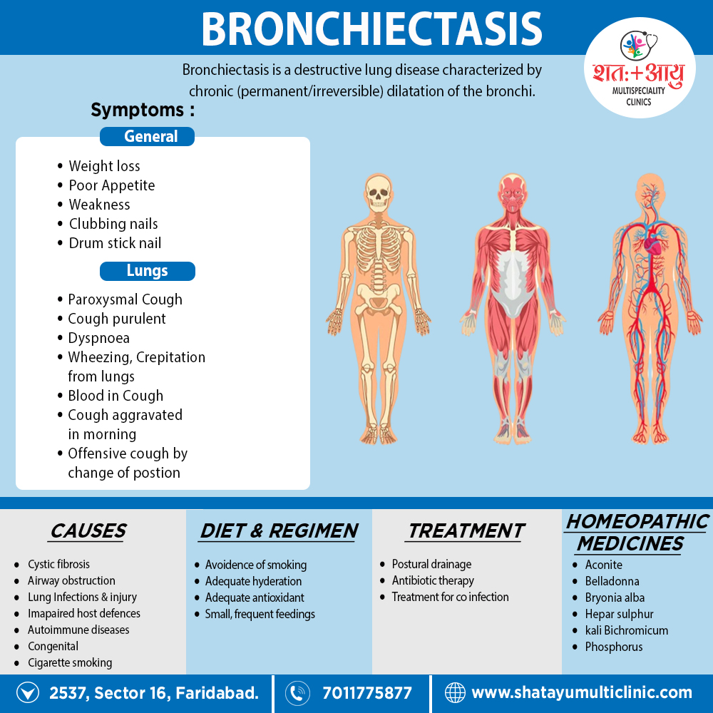
Pathophysiology

The diagnostic feature of bronchiectasis dilate bronchi. The Reid classification differentiates between pathological and radiological appearances of bronchiectasis.

Cylindrical bronchiectasis i.e.

  1. Bronchial dilatation is mild and the bronchi retain their regular relatively straight outline.
  2. Varicose bronchiectasis
  3. Bronchial dilatation is greater and local constrictions are present, giving the airway an irregular appearance.

Saccular/cystic bronchiectasis i.e.

–is the most severe form and characterize by large areas of distal ‘grape-like’ bronchial dilatation and loss of bronchial subdivision.

Atelectatic bronchiectasis i.e.

-is a localized form related to proximal bronchial distortion or occlusion.

Sign & Symptoms

Bronchitis i.e.

Attacks of recurrent bronchitis, more common in winter. Clubbing of fingers diagnostic.

Hemorrhagic (bronchiectasis sicca) –

Recurrent hemoptysis with good health in-between, or attacks of bronchitis.

Suppurative –

For instances; Chronic cough, copious purulent expectoration, general toxemia, clubbing of fingers varying from slight parrot beak curvature of finger nails to bulbous drum stick enlargement (pulmonary osteoarthropathy).

During exacerbations, Dyspnoea and wheezing occur in 75% of patients and pleuritic chest pain in 50%. Pleuritic chest pain cause by distended peripheral airways and inflammation adjacent to the visceral pleura. Pyrexia rare during exacerbations; if it is present, pneumonia should exclud.

With relatively rapid onset –

Symptoms developing with comparative suddenness, as a sequel to partial bronchial obstruction by a foreign body or after anesthesia. In early stages paroxysmal cough with occasional offensive sputum which may provoke by change of position. Later large amounts of foetid sputum. Signs mostly limited to auscultation and depend on the size of the affected bronchi, patency of the airways and viscosity of secretions. There may signs of (a) bronchitis, or (b) fibrosis, or (c) consolidation, or (d) collapse, or (e) of cavitation’s.

Early stages –

Fine crackles or sticky rhonchi and slight alteration in character of breath sounds.

Late stages –

Bronchial breathing, coarse crept and perhaps signs of a cavity. Changing character of physical signs after a long bout of cough when air entry may become louder and bronchial in character or from day to day. Sharp metallic or “leathery” rales characteristic. Recurrent pneumonia in the same area of the lung classically associate with bronchiectasis. [1]

Diagnosis

SPUTUM EXAMINATION –

To exclude diagnosis of underlying pulmonary tuberculosis. Culture and sensitivity of infecting organism as a guide to chemotherapy.

RADIOLOGY:

Sinus radiographs – for chronic sinusitis.

Chest radiography – for changes suggestive or associated with the diagnosis.

Collapse (either segmental or lobar)

Crowding of pulmonary vessels, indicating area of damage or consolidation that may become infected. • ‘Tramline shadows’ suggesting bronchial wall oedema.

Cystic lesions suggesting saccular bronchiectasis

Evidence of previous tuberculosis.

Evidence of previous heart surgery.

HRCT–

High-resolution CT has replaced bronchography and is the preferred investigation. CT findings include the following: – The ‘signet ring’ sign (end-on dilated bronchi that are larger than the accompanying pulmonary artery) seen in all form of bronchiectasis

Tramlines (in other words, non-tapering bronchi) seen in cylindrical bronchiectasis – Varicose bronchiectasis has a beaded (tree-imbued) appearance – ‘Cysts’ with air-fluid levels in dilated bronchi characteristic of cystic bronchiectasis SPIRAL CT may aid diagnosis by reducing motion artefact and identifying subtle bronchiectatic changes.

SPECIAL INVESTIGATIONS:

may indicate the cause of bronchiectasis in some patients.

Immunoglobulins –

Most patients with bronchiectasis have raised plasma concentrations of immunoglobulins (especially IgA).

Aspergillus precipitins –

Recurrent mucus plugging of major airways and development of proximal bronchiectasis occurs in allergic bronchopulmonary aspergillosis.

Barium studies –

For gastro-esophageal reflux, since association of basal bronchiectasis is well recognized.

a1-antitrypsin deficiency associate with premature pulmonary emphysema though rarely in association with bronchiectasis.

Detection of cystic fibrosis –

By sweat test and/or genetic analysis

Treatment

Postural drainage–

Since bronchiectasis usually affects the lower lobes the prone position with the head tilted downwards is the most useful. Deep breathing or coughing, assisted by percussion of the affected part (clapping) helps to dislodge the secretions. It should carried out at least twice daily for 15–20 minutes at a time. Expectoration facilitate by cough mixtures, or better inhalation of nebulized bronchodilator isoproterenol followed by inhalation of nebulized water or steam, followed by postural drainage. This should done on arising in the morning since the patient with bronchiectasis tends to pool secretions during the night. [1]

Antibiotic therapy

Antibiotics can delay progression in patients with cystic fibrosis. Antibiotics penetration into the bronchial mucosa and secretions impair and the dose needs to be higher and duration longer. H. influenzae, H. parainfluenza, Str. pneumonia and Moraxella catarrhalis are the most common organisms in exacerbations,

  • β-lactam antibiotics (e.g. amoxicillin 500 mg t.d.s. for 10–14 days) remain the first line therapy.
  • Alternatives include quinolones and macrolides. Chronic colonization with Ps. aeruginosa is common in severe disease.
  • Choice of antibiotic guide by sensitivities, but oral quinolones (e.g. ciprofloxacin 500 mg b.d. for 2 weeks) is the first time therapy. Development of resistance is common, and iv antibiotics e.g. ceftadizime 2 g t.d.s. for 14 days, gentamicin (in accordance with plasma levels), and piperacillin 4.5 g t.d.s. for 14 days require. Nebulized antibiotics allow maximal concentrations of antibiotics in the airways with reduced systemic effects. Amoxicillin 500 mg b.d., gentamicin 80 mg b.d., and tobramycin solution for inhalation 300 mg b.d. can be used.
Acute exacerbations –

Hospitalization in all severely ill patients, when chest pain limits coughing and sputum clearance (this increases risk of pneumonia) and iv antibiotics are required.

Prophylactic therapy –

One guideline for therapy is when there are exacerbations every 2 months that prevent participation in normal activities of 2 weeks or more during the exacerbation. Continuous therapy considere in patients who persistently expectorate purulent sputum. Aggressive treatment warrant because persistent infection causes tissue destruction. Bilateral lung transplantation has use when respiratory failure develops despite optimal medical management.

Intravenous immunoglobulin replacement

Intravenous immunoglobulin replacement thought to be effective in pan hypogammaglobulinemia, but given only to patients with established, widespread lung damage.

α-antitrypsin replacement therapy by inhaled route has the potential to neutralize damaging processes that are released as part of the airways inflammatory response.

General supportive treatment – (a) Adequate nutrition. (b) Eradication of chronic nocturnal post-nasal discharge and treatment of sinusitis. (c) Avoidance of smoking. (d) Adequate hydration

Complications of bronchiectasis

Pulmonary – recurrent pneumonia, lung abscess, haemoptysis

Pleural – pleurisy, pleural effusion or empyema Corpulmonale Cerebral abscess Amyloidosis Seronegative arthropathy. [1]

Homeopathic Treatment

Homeopathy treats the person as a whole. It means that homeopathic treatment focuses on the patient as a person, as well as his pathological condition. The homeopathic medicines are selected after a full individualizing examination and case-analysis,

which includes

  • the medical history of the patient,
  • physical and mental constitution,
  • family history,
  • presenting symptoms,
  • underlying pathology,
  • possible causative factors etc.

The disease diagnosis is important but in homeopathy, the cause of disease is not just probed to the level of bacteria and viruses. Other factors like mental, emotional and physical stress that could predispose a person to illness are also looked for. No a days, even modern medicine also considers a large number of diseases as psychosomatic. The correct homeopathy remedy tries to correct this disease predisposition. The focus is not on curing the disease but to cure the person who is sick, to restore the health. If a disease pathology is not very advanced, homeopathy remedies do give a hope for cure but even in incurable cases, the quality of life can be greatly improved with homeopathic medicines.

The homeopathic remedies

The homeopathic remedies (medicines) given below indicate the therapeutic affinity but this is not a complete and definite guide to the homeopathy treatment of this condition. The symptoms listed against each homeopathic remedy may not be directly related to this disease because in homeopathy general symptoms and constitutional indications are also taken into account for selecting a remedy.


ACONITUM NAPELLUS:

The first remedy that is often indicated at the onset of the infection. It is effective only within the first twenty four hours. Useful when there is a short, dry cough, a temperature and an irritation of the throat, chest and trachea. The cause is usually exposure to cold. There is a chilly restlessness, anxiety and a full, bounding pulse, with general weakness.

ANTIMONIUM TARTARICUM:

Useful when there is a accumulation of considerable loose, rattling, moist mucus in the chest. For children breathing and a loose cough, but little phlegm is expectorated. There may be vomiting and laboured breathing. Pronounced physical and mental exhaustion.

BELLADONNA:

A high temperature, dry cough, a pounding, pulsating headache and a flushed face with a dry, hot skin. The cough is worse at night and when lying down.[2]

BRYONIA ALBA:

A painful, violent, dry, stitching cough with a headache and pain in the chest wall. Worse with coughing and better when supporting the area with both hands. The expectoration is yellow and often blood streaked. The cough is worse after meals. Patient drinks large quantities of fluids at long intervals.

KALI BICHROMIUM:

For less acute cases. The phlegm is tough, thick and stringy and cannot easily be expectorated. There is a tightness of the chest and a troublesome cough.

Worse in the evenings. (about 4 pm to 5 pm).

PHOSPHORUS:

Useful for tall, pale patients with little natural resistance. They are bright-eyed, very anxious and need constant reassurance. Breathing is often difficult with wheezing; with a dry, tickling cough and a sore chest. Worse when talking and in fresh air.

BACILLINUM:

Repeated infections.[2]

Diet & Regimen

  • Use a diet with 1.0–1.25 g protein/kg and sufficient calories to meet elevated metabolic requirements appropriate for age and sex.
  • Small, frequent feedings may better tolerate.
  • Fluid intake of 2–3 L daily may offer, unless contraindicated.
  • Intravenous fat emulsions may indicate (eicosanoids are inflammatory modulators, and thromboxane and leukotrienes tend to be potent mediators of inflammation). Omega-3 fatty acids should enhance in the oral diet by including salmon, tuna, sardines, walnuts, and flaxseed. Supplements may also useful.
  • Adequate antioxidant use with vitamins C and E and selenium may beneficial. Ensure adequate potassium intake, depending on medications used.[3]

FAQs

Frequently Asked Question

What is Bronchiectasis?

Bronchiectasis is a destructive lung disease characterized by chronic (permanent/irreversible) dilatation of the bronchi associated with persistent though variable inflammatory process in the lungs.

Homeopathic Medicines used by Homeopathic Doctors in treatment of Bronchiectasis?

  • Aconitum Napellus
  • Antim Tart
  • Belladonna
  • Bryonia
  • Kali Bichromicum
  • Bacillinum

What are the symptoms of Bronchiectasis?

  • Bronchitis- Clubbing of fingers
  • Hemorrhagic- Hemoptysis
  • Suppurative- Chronic cough, Copious purulent expectoration
  • Dyspnea and wheezing
  • Paroxysmal cough

What are the causes of Bronchiectasis?

  • Cystic fibrosis
  • Airway obstruction
  • Lung infections
  • Impaired host defenses
  • Aspergillosis
  • Autoimmune diseases
  • Lung injury
  • Congenital
  • Cigarette smoking

References:

  1. Golwalla’s MEDICINE for Students 25th Edition
  2. Homoeopathic Body-System Prescribing – A Practical Workbook of Sector Remedies
  3. Nutrition and Diagnosis-Related Care (Nutrition and Diagnosis-Related Care (Escott-Stump)